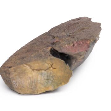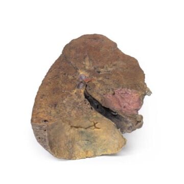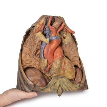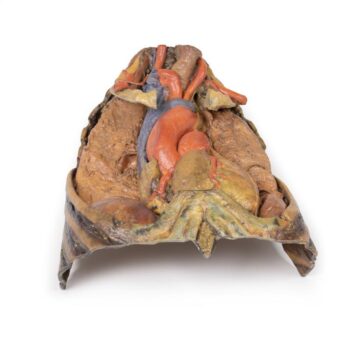



Features
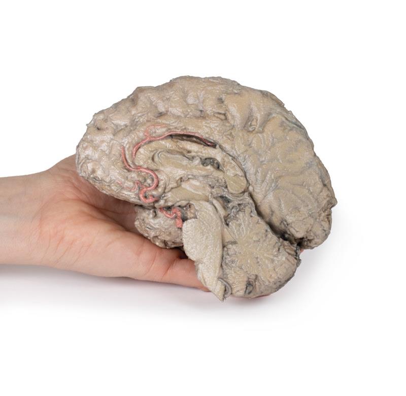
The Brain Hemisection is a 3D printed anatomical model designed for medical educators, clinicians, and students who need precise brain anatomy representation. It offers detailed views of preserved right brain structures and midline features, providing a reliable resource for teaching and advanced study. Choose this model to eliminate the complexities and costs of human cadaveric specimens while enjoying clinical-grade accuracy and immediate availability.
See the Brain as Never Before with Clinical-Grade 3D Accuracy
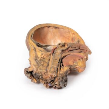
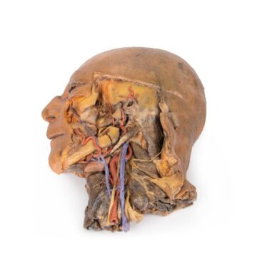
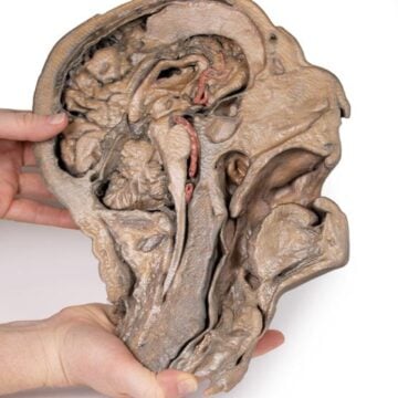
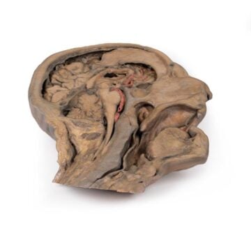
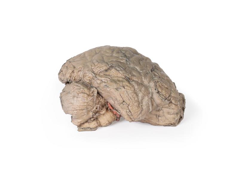
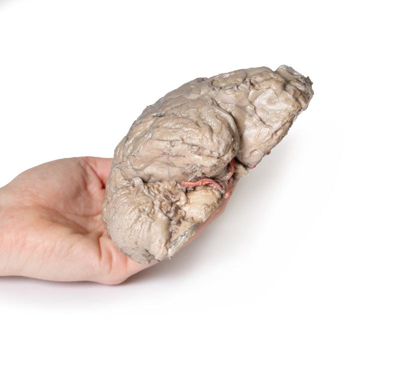
This Brain Hemisection delivers a complete midsagittal cut that preserves right-sided cerebral and cerebellar anatomy, deep brain structures, and key neurovascular features. In lateral view, you can observe intact arachnoid mater covering both the right cerebral and cerebellar hemispheres. The model reveals midline areas from the cerebrum to the medulla oblongata, with central detail including the third ventricle, septum pellucidum, right mamillary body, and optic tract. Vibrant false-coloured arteries make neurovascular pathways instantly recognizable, supporting in-depth learning and demonstration. Avoid ethical and logistical issues associated with cadaver use and benefit from a durable, accessible, and highly accurate learning tool.
Features and Benefits
- True-to-specimen 3D printed from real human anatomy
- Preserves right cerebral and cerebellar hemispheres in the arachnoid mater
- Midline view retains cerebrum to medulla oblongata structures
- Opened third ventricle with visible septum pellucidum
- Right mamillary body and optic tract displayed
- Posterior view includes cerebral aqueduct from midbrain to fourth ventricle
- Shows tentorium cerebelli and cerebellar cortex forming arbor vitae
- False-coloured arterial branches highlight major neurovascular pathways
- Anterior cerebral artery, middle cerebral artery, and superior cerebellar artery pathways detailed
- Reproducible, consistent and easy to customize
- No ethical collection, handling or storage issues
- Lower cost and fast availability compared to cadaveric specimens
Indications for Use
- Medical and allied health education
- Anatomy teaching in universities and clinical settings
- Surgical or neuroscientific training
- Patient communication and demonstration
- Standardized assessment and examinations
Size Guide
- One standard clinical-education size only – scaled to life-size brain dimensions for accurate representation and compatibility with teaching setups