
Features
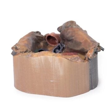
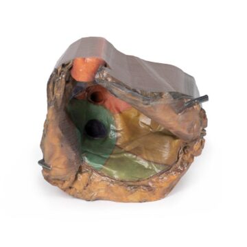
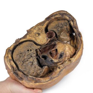
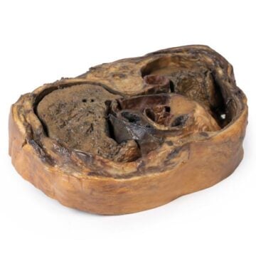
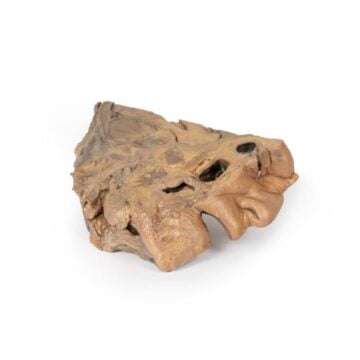
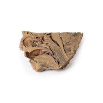
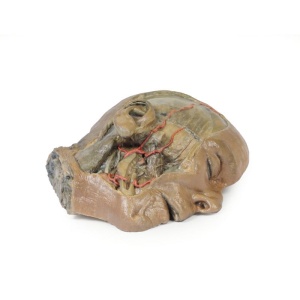
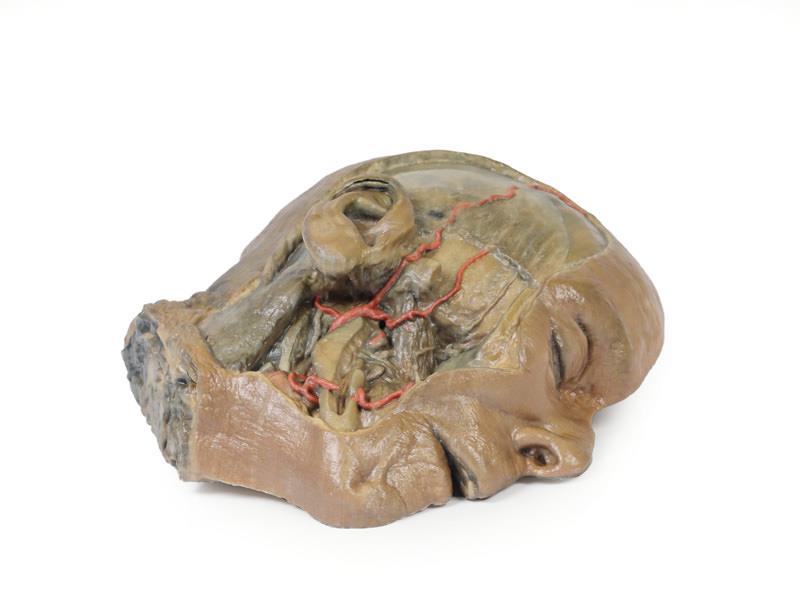
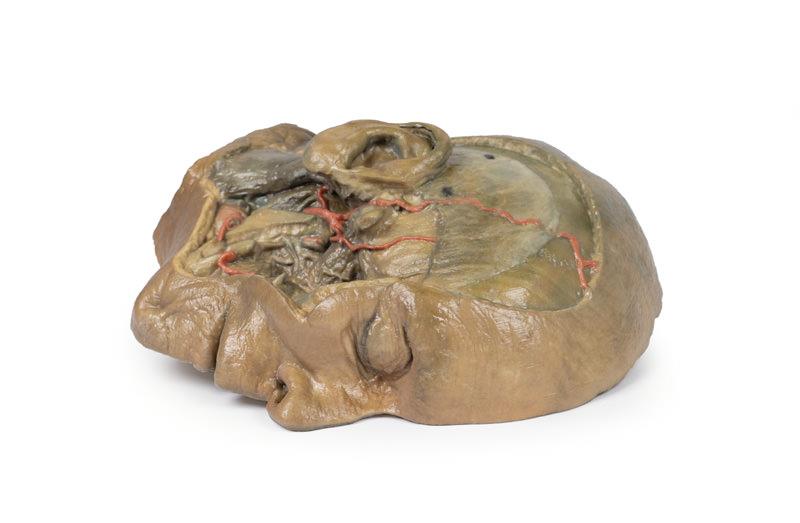
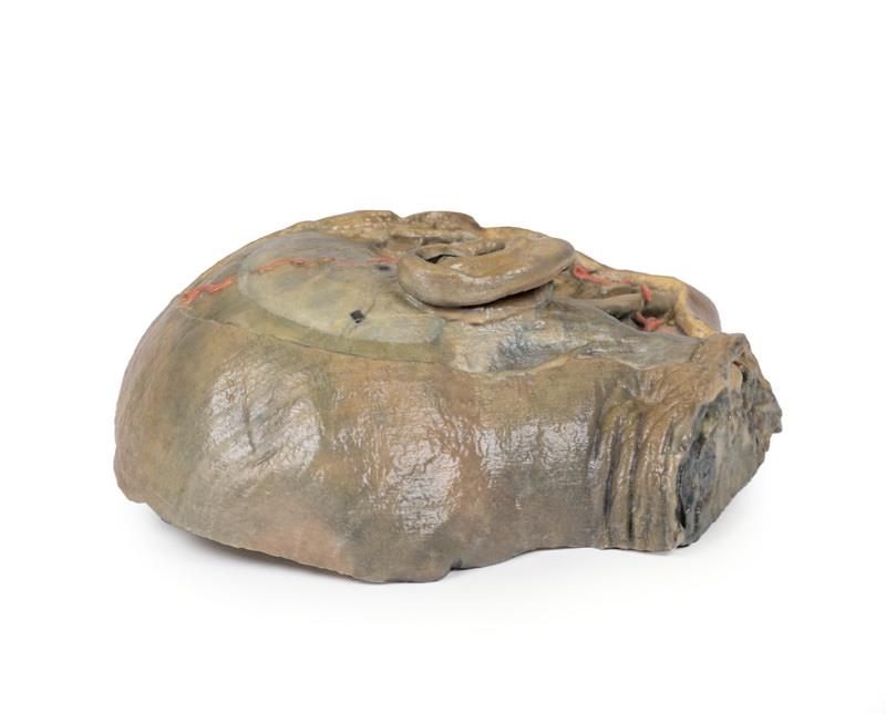
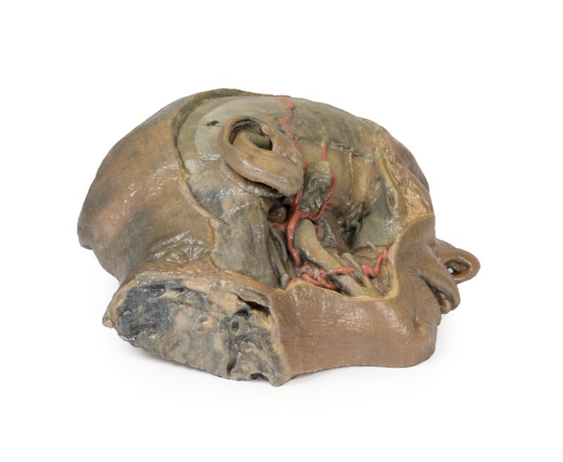
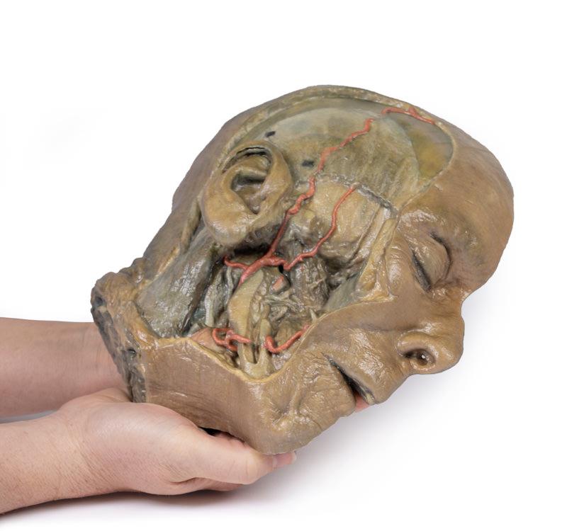
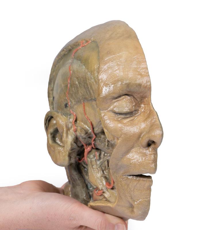
The Sagittal Section of head with infratemporal Fossa Dissection is a highly detailed 3D anatomical model designed for medical educators, clinicians, and students. It enables advanced study of head and neck structures through its comprehensive midline section and intricate infratemporal fossa dissection. This model ensures a clear, hands-on understanding of complex cranial and facial anatomy, making it an indispensable resource for anatomy education and clinical demonstration.
Unlock Deeper Anatomical Insight with the Sagittal Section of head with infratemporal Fossa Dissection
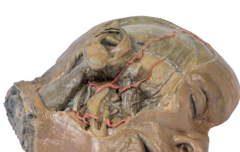
This 3D printed model provides unmatched visibility into head, neck, and infratemporal fossa anatomy. It presents a midsagittal head section with preserved endocranial contents, nasal and oral cavities, and pharynx down to the laryngeal cartilages. The infratemporal fossa features detailed dissections, showing arterial, venous, neural, and glandular structures not visible in traditional models. Use this clinical-grade teaching tool to enhance demonstrations, surgical planning, or anatomical education with a level of realism and specificity that 2D diagrams cannot achieve.
Features and Benefits
- Combined midsagittal and infratemporal fossa dissection in a single 3D model
- Preserved endocranial contents, nasal and oral cavities, and pharynx to laryngeal cartilage level
- Nearly intact nasal cavity with ethmoid air cells and large individual sphenoid sinus
- Visible lateral ventricle with partial fourth ventricle and separated cingulate gyrus and corpus callosum
- Exposed optic tract, pituitary gland, colliculi, cerebellar peduncle, and medulla-spinal cord transition
- Detailed view of tentorium cerebelli and visible cerebral/cerebellar arteries
- Extensive scalp dissection highlighting the posterior auricular nerve and temporal arteries
- Deep infratemporal fossa view with dissected mandibular structures, arteries, veins, and nerves
- Clear identification of external and maxillary arteries, deep temporal arteries, retromandibular vein, facial artery, internal jugular vein, lingual nerve, and parotid duct
- Clinical-grade build suited for repeated hands-on use
Indications for Use
- Anatomy education in medical and dental schools
- Clinical demonstration for healthcare professionals
- Surgical planning and preoperative training
- Patient education and consultation support
Size Guide
- One size only – 3D printed life-size human head and neck section
- Replicates anatomical proportions based on adult human specimen
- Contact supplier for exact measurements or custom model requests