



Features
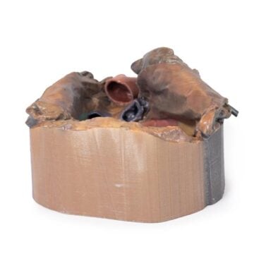
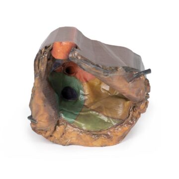
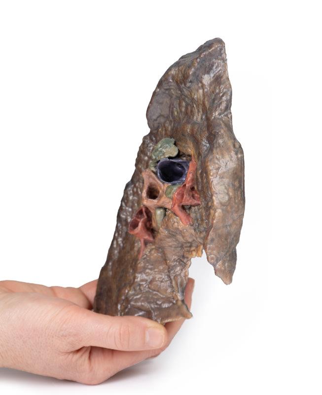
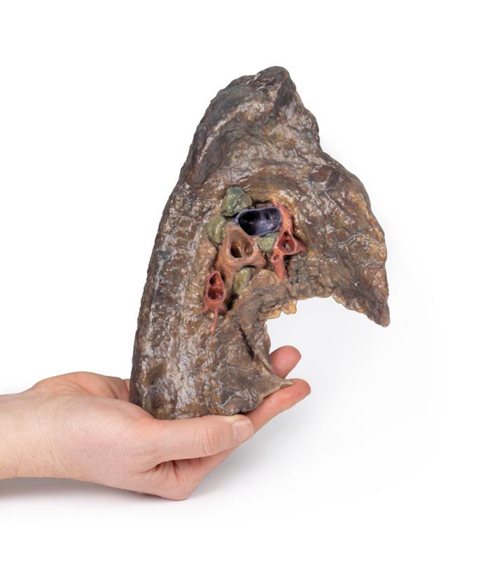
The Hilum of the left lung is a premium 3D printed anatomical model designed for medical educators, students, and healthcare professionals. It provides an accurate, enlarged view of the lung’s vital entry and exit point, helping users master the complexities of pulmonary anatomy. Choose this detailed replica to enhance anatomical teaching, clinical training, and patient education in any academic or clinical setting.
Unlock a Precise Understanding of Pulmonary Anatomy with the Hilum of the left lung
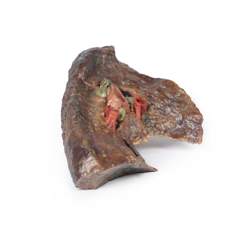
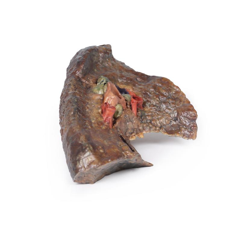
This advanced 3D printed model highlights the comprehensive structure of the Hilum of the left lung. It clearly presents the junction where the visceral and parietal pleura meet and demonstrates the lung’s only connection with the rest of the body through the pulmonary ligament. The model includes the pulmonary artery, superior and inferior pulmonary veins, main bronchi, nerves, and lymphatics, all arranged in anatomical accuracy. Meticulously sectioned in a sagittal plane along the cardiac notch, it reveals the spatial relationships and detailed vascular and airway pathways found in the hilum. The lifelike architecture, visible fissures, vascular impressions, and lymph node clusters empower users to study and teach the region’s complexity.
Key Features:
Hilum Anatomy:
– Entry/exit point for the pulmonary artery, superior and inferior pulmonary veins, main bronchus, lymphatics, and nerves.
– Shows the meeting point of visceral and parietal pleura, forming the pulmonary ligament—the lung’s sole anatomical connection to the body.
Pulmonary Circulation:
– Pulmonary artery (superior in position) carries deoxygenated blood from the heart.
– Pulmonary veins (anterior and inferior) return oxygenated blood to the heart.
Bronchial Structure:
Left main bronchus and its lobar branches are visible, located posteriorly within the hilum.
Additional Views:
– Oblique fissure along the lateral lung surface.
– Diaphragmatic surface at the base; costal visceral surface posteriorly.
– Pulmonary lymph nodes surrounding the hilum, both medially and laterally.
Indications for Use
- Medical and university anatomy education
- Clinical demonstration to healthcare students and professionals
- Patient education for pulmonary procedures
- Reference for surgical planning and anatomy review
Size Guide
- Scaled for anatomical accuracy, reflects dimensions of the left lung hilum in adult humans
- Model dimensions match standard anatomical references for educational effectiveness