Features
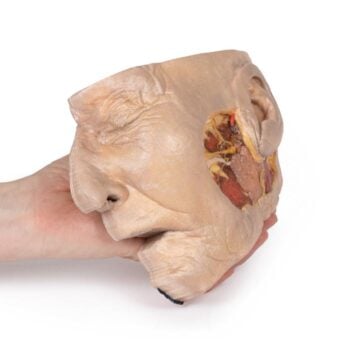
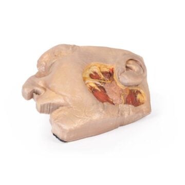
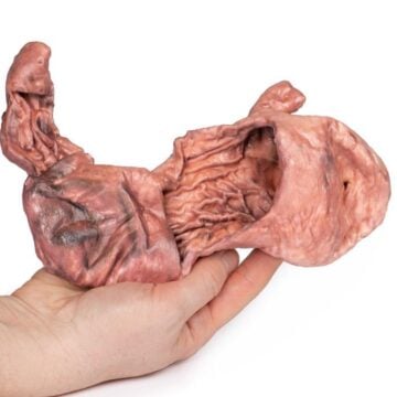
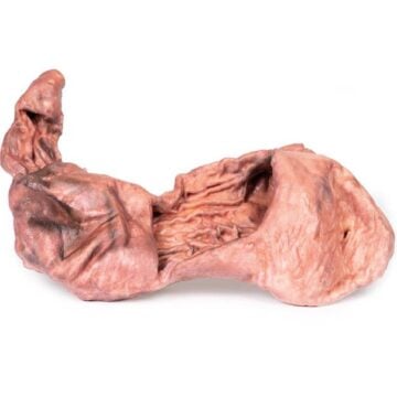
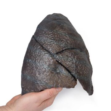
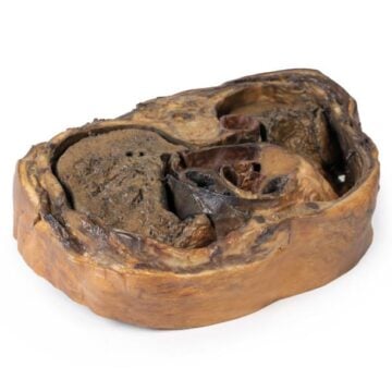
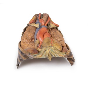
The Thorax with heart and vessels is a high-fidelity 3D printed anatomical model crafted for advanced medical education and clinical demonstration. It provides students and healthcare professionals with an exceptional resource to explore intricate thoracic structures. You gain access to detailed observation of the heart, major vessels, nerves, ribs, and musculature, making this model invaluable for anatomy instruction and procedural training.
Reveal True-to-Life Thoracic Anatomy with the Thorax with heart and vessels
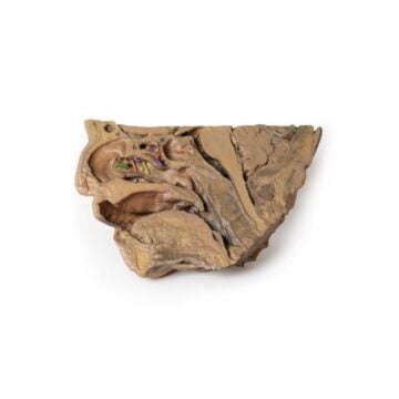
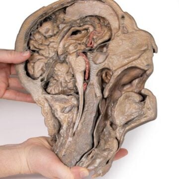
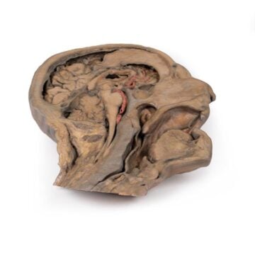
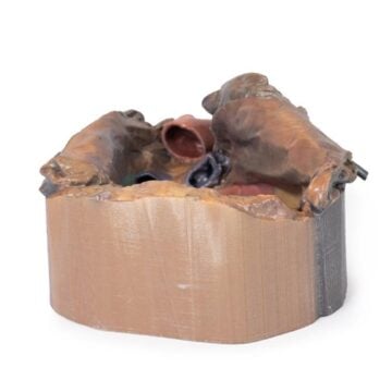
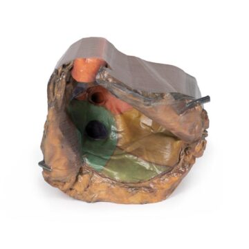
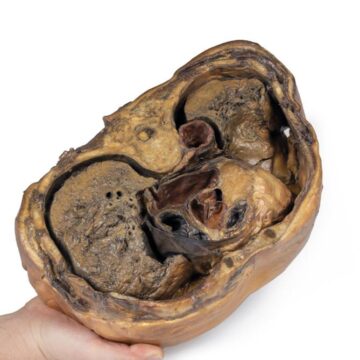
This premium model showcases detailed reproductions of thoracic organs and major vascular pathways. You can study the trachea with thick cartilage, observe both right and left subclavian arteries and their branches, and trace nerve pathways like the brachial plexus and phrenic nerves. Dissection exposes key anatomic views, including the left vagus and recurrent laryngeal nerves, the pulmonary trunk, and the arch of the aorta. The model captures rib eight through twelve and right and left hemidiaphragms, delivering realistic teaching and assessment experiences.
Features and Benefits
- 3D printed construction ensures anatomical precision and clinical-grade detail
- Displays thoracic aperture, heart, major vessels, and complete mediastinum structures
- Shows both subclavian arteries with visible branches and common carotid arteries
- Highlights brachial plexus roots, trunks, and smaller nerve branches
- Exhibits trachea, rib one, and external intercostal muscle anatomy
- Reveals both right and left phrenic and vagus nerves with associated pathways
- Presents major vessels including arch of aorta, pulmonary trunk, and superior vena cava
- Illustrates right and left hemidiaphragms with relevant musculature and ribs eight-twelve
- Clinical-grade, education-standard model for reliable training and demonstration
Indications for Use
- Medical and anatomy education for students and healthcare professionals
- Clinical demonstration and procedural training
- Specialist instruction in thoracic structures, vessels, and nerves
- Ideal for universities, hospitals, and research institutions
Size Guide
- One universal size – full-scale reproduction of thorax region
- Includes anterior, superior, and inferior thoracic perspectives
- Model dimensions are true to adult human anatomy for accurate visualization