Features
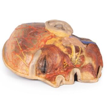
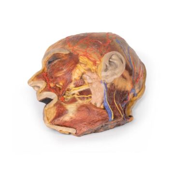
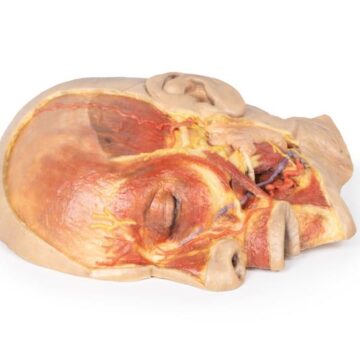
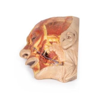
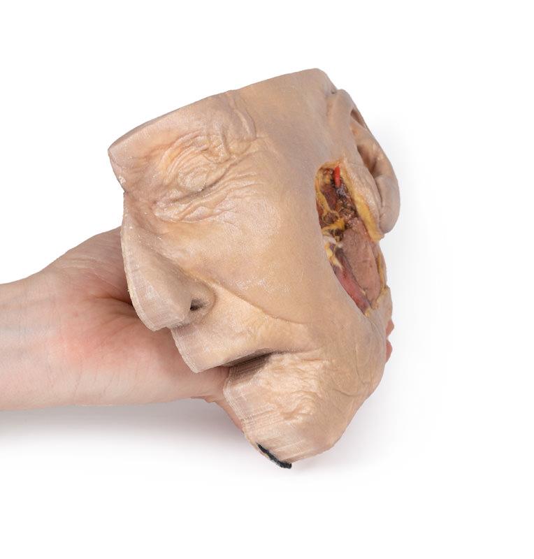
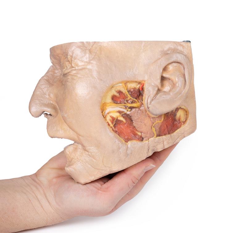
The Parotid Gland and Facial Nerve dissection is a 3D printed anatomical model designed for medical professionals, educators, and students. It provides unmatched clarity in demonstrating the intricate relationship between the parotid gland, facial nerve, and surrounding structures. Choose this model for its clinical accuracy and practical value in advanced surgical planning and anatomical education.
Revolutionize Learning and Surgery Preparation with an Unmatched Anatomical Reference
This model opens a realistic window into the lateral face, revealing detailed anatomy significant in Mohs surgery, skin cancer management, and reconstructive procedures. The Parotid Gland and Facial Nerve dissection highlights precise relationships among neurovascular structures and surface anatomy, helping users visualize the facial nerve’s terminal branches, the parotid duct, the parotid gland, and the great auricular nerve. Its lifelike presentation supports confident procedure rehearsal and deepens anatomical understanding for surgeons, trainees, and educators. The sturdy, clinical-grade construction ensures durability and repeated use in teaching environments.
Features and Benefits
- True-to-life 3D printed model with superficial dissection window
- Clear presentation of the parotid gland’s relation to facial anatomy
- Exposes and details superficial temporal artery, facial nerve branches, and parotid duct
- Displays the great auricular nerve near the parotid gland and sternocleidomastoid
- Ideal for demonstrating anatomical landmarks vital to advanced surgeries
- Built from clinical-grade materials for professional education
Indications for Use
- Preparation and planning for Mohs surgery
- Skin cancer surgical education
- Plastic and reconstructive surgery training
- Medical anatomy teaching and demonstration
Size Guide
- Standard model – anatomical region extends from the zygomatic arch to the angle of the mandible, and from the anterior margin of the masseter to the origin of the sternocleidomastoid