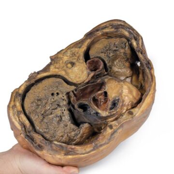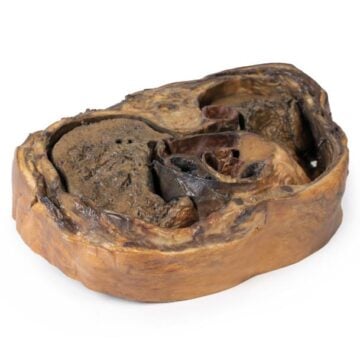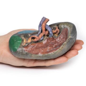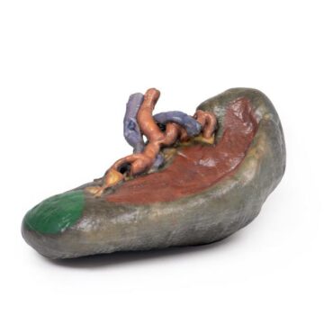


Features
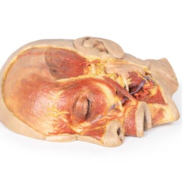
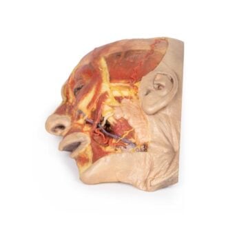
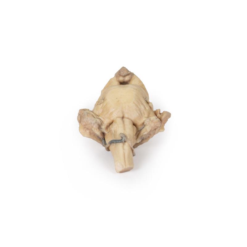
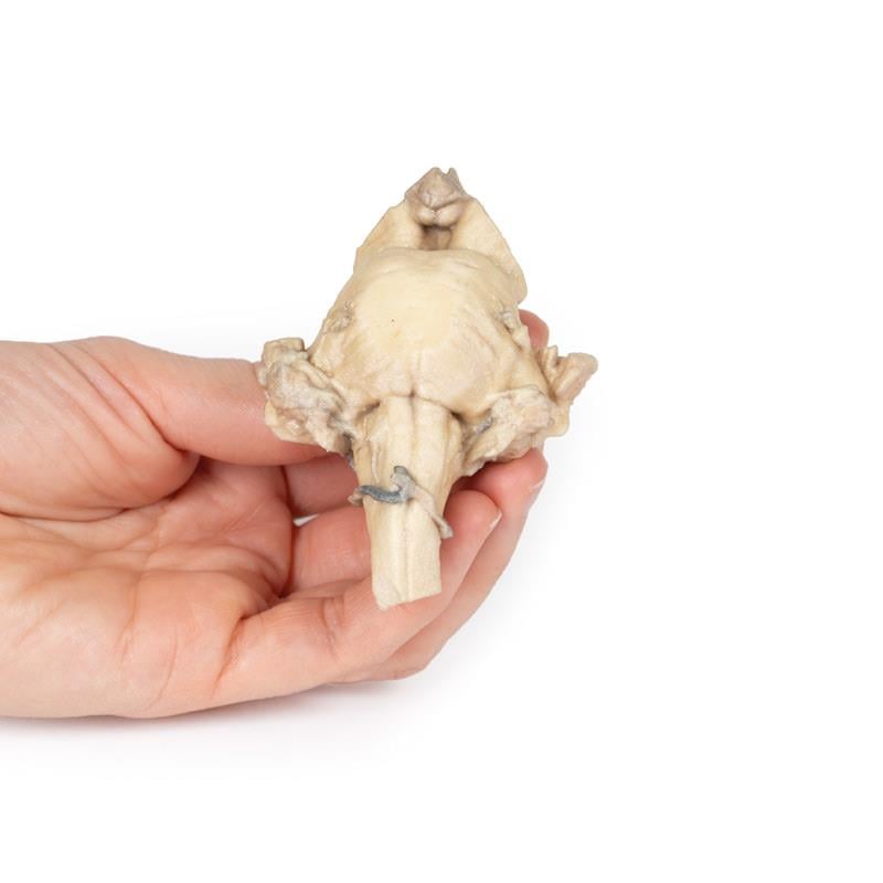
The Brain Stem, isolated anatomy from midbrain to medulla oblongata is a 3D printed anatomical model designed for educators, clinicians, and students. It provides a realistic and detailed depiction of the brainstem, helping users visualize key structures and understand complex neuroanatomy. Choose this model to avoid the limitations of cadaveric specimens and ensure precise, consistent educational value.
Enhance Neuroanatomy Education with Accurate 3D Brainstem Representation
Dive into unparalleled anatomical realism with the Brain Stem, isolated anatomy from midbrain to medulla oblongata. This clinical-grade, 3D printed model shows the brainstem from the midbrain to the medulla oblongata. The anterior view displays the mamillary bodies sitting between the cerebral peduncles and the pineal gland. Posterior highlights include the corpora quadrigemina and clear superior cerebellar peduncles. The cerebellum is removed, exposing the cross-sections of cerebellar peduncles and the fourth ventricle. The preserved pons includes the trigeminal nerve origin, while the medulla features prominent pyramids and olives. You avoid cumbersome cadaver requirements while gaining reproducible, customizable, and affordable anatomical accuracy. This model sets a new standard for medical training and education.
Features and Benefits
- 3D printed from real human anatomical data
- Clinical-grade accuracy for education
- Shows full brainstem from midbrain to medulla oblongata
- Displays anterior and posterior landmarks, including mamillary bodies, pineal gland, corpora quadrigemina
- Removable cerebellum for detailed examination of peduncles and fourth ventricle
- Pons with preserved trigeminal nerve origin
- Visible pyramids and olives on the medulla
- Lower cost and faster availability than cadaveric specimens
- No ethical or handling concerns
- Reproducible for standardized education
- Customization options for enlargements, structure highlights, and cutaway views
Indications for Use
- Anatomy education for students, clinicians, and educators
- Medical training institutions seeking lifelike models
- Demonstration of neuroanatomical relationships in the brainstem
- Replacement or supplement for cadaveric specimen teaching
- Simulation of specific neurological structures for advanced study
Size Guide
- One standard model size – full brainstem from midbrain to medulla oblongata as per anatomical dimensions
- No alternative sizes available – model designed to true anatomical scale