Features
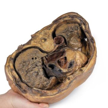
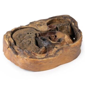
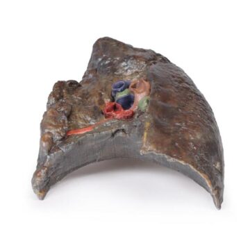
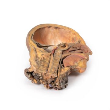
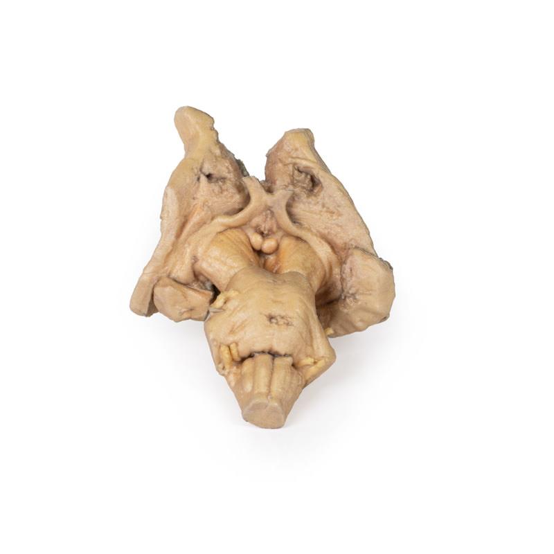
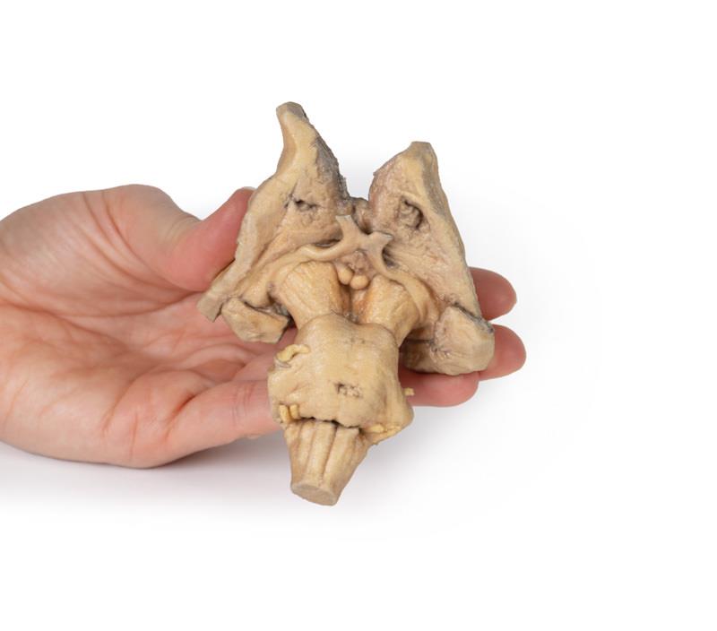
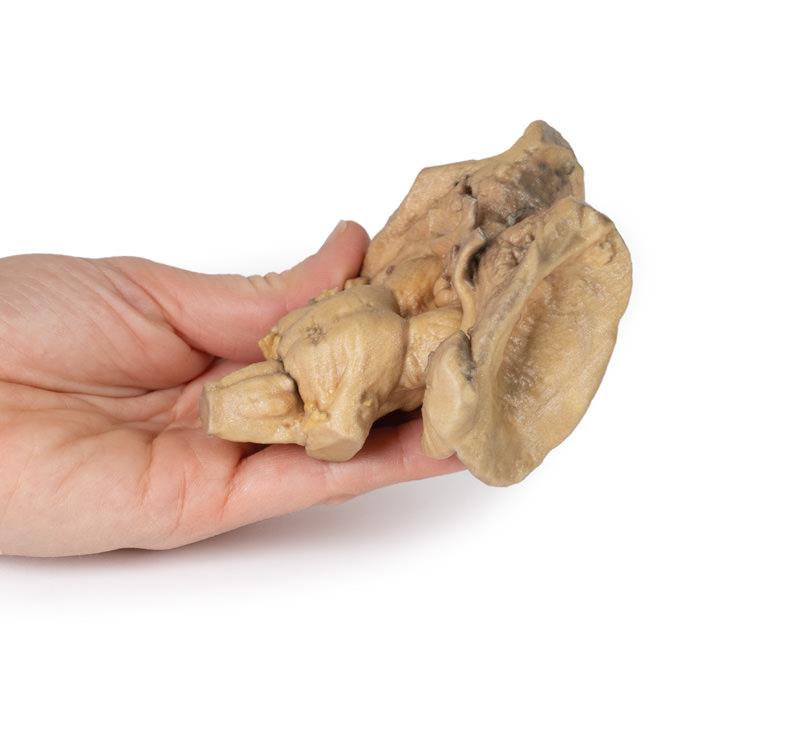
The Brain stem, deep cerebral and diencephalic structures is a 3D printed anatomical model designed for medical educators, students, and clinicians who demand the most accurate, detailed representation of deep brain anatomy. This model solves the need for highly realistic, cadaver-derived structure without the storage and ethical complexities of human specimens. Choose this product for uncompromising educational value, reproducibility, and customization to suit your learning goals.
Experience Unmatched Anatomical Detail and Clarity in Neuroanatomy Education
Dive into advanced neuroanatomy with the Brain stem, deep cerebral and diencephalic structures model. Featuring meticulously preserved cerebral and diencephalic structures down to the proximal medulla oblongata, this model showcases authentic detail on every aspect. Discover the lentiform nucleus, corona radiata, caudate nucleus, thalami, and amygdaloid bodies, all faithfully printed from real human specimens. Explore anterior structures such as cerebral peduncles and optic nerves, as well as posterior and inferior features like the superior and inferior colliculi, fourth ventricle, and cranial nerve origins. Elevate your teaching and learning experience with a reusable model that removes the logistical and ethical hurdles of cadaver specimens while delivering consistent quality.
Features and Benefits
- 3D printed from actual human brain specimens for maximum anatomical accuracy
- Preserves deep cerebral and diencephalic structures to proximal medulla oblongata
- Presents lentiform nucleus and corona radiata on right side
- Shows caudate nucleus head/body, thalami, amygdaloid bodies, and third ventricle
- Detailed anterior with cerebral peduncles, optic nerves, and mammillary bodies
- Displays interpeduncular region, pons, and origins of cranial nerves V, VII, VIII
- Features prominent pyramids and olives on medulla oblongata
- Exposes superior/inferior colliculi, fourth ventricle, and rhomboid fossa floor structures
- Avoids ethical complications and storage needs of cadaveric specimens
- Cost-effective, reproducible, and consistent for standardized education
- Customizable—highlight, enlarge, or section specific anatomical areas as needed
Indications for Use
- Ideal for teaching neuroanatomy in universities and medical schools
- Essential for clinical demonstration and anatomical reference for neurologists and neurosurgeons
- Training and assessment in anatomy for allied health education
- Valuable for research, development, and simulation in medical device studies
Size Guide
- Single model representing authentic human brain structures—actual anatomical proportions
- No alternative sizes or variants available