



Features
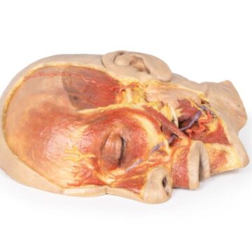
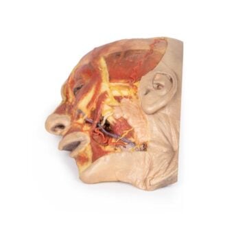
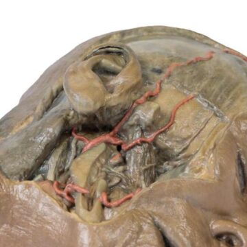
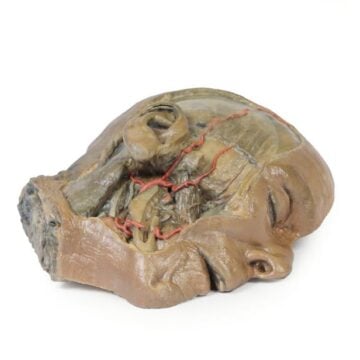
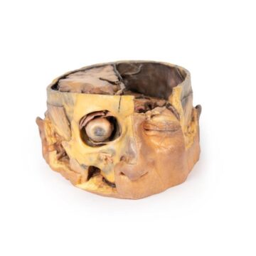
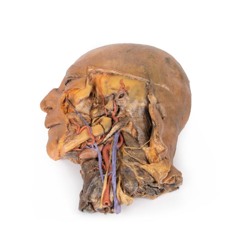
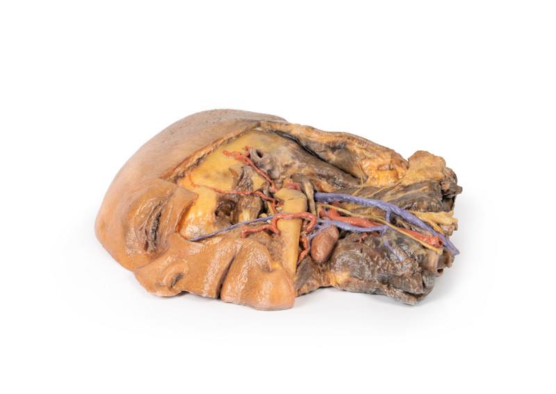
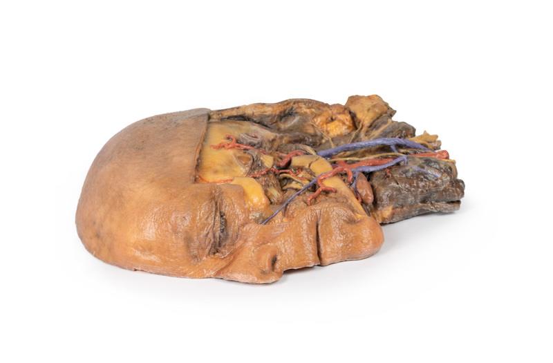
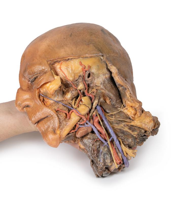
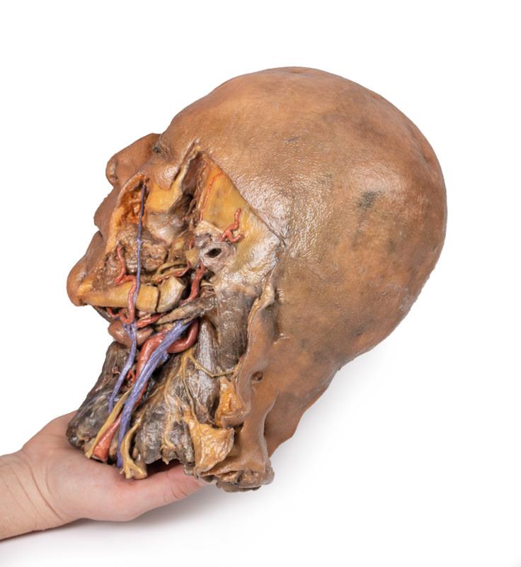
The Sagittal Section of Head and Neck with Infratemporal Fossa and Carotid Sheath Dissection is a clinically accurate 3D printed anatomical model built for educators, students, and healthcare professionals. It delivers a comprehensive view of the head and neck by revealing vital endocranial and neck structures with lifelike clarity. Select this model to elevate anatomical understanding and bridge the gap between theory and real-world dissection.
Unmatched Visualization for Comprehensive Anatomical Education
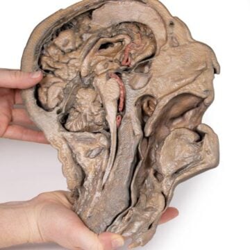
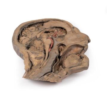
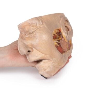
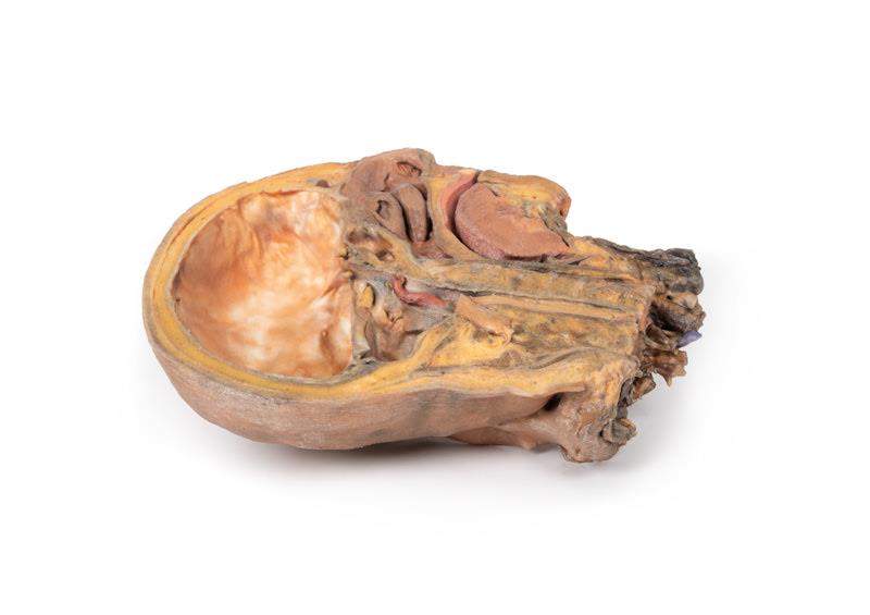
Elevate your teaching and study sessions with the Sagittal Section of Head and Neck with Infratemporal Fossa and Carotid Sheath Dissection. This model provides a detailed midsagittal and lateral dissection, exposing the endocranial cavity without the brain and key neck anatomy. Clear visual access to dura mater, tentorium cerebelli, cranial nerves, pituitary gland, vessels, and the infratemporal fossa supports advanced learning and instruction. The precision and clinical-grade quality ensure reliable use in university, clinical, or professional settings.
Features and Benefits
- True-to-life 3D printed model
- Midsagittal section with brain removal for enhanced view of dura mater and tentorium cerebelli
- Visible cranial nerves including optic, oculomotor, trigeminal, abducens, facial, and vestibulocochlear
- Cross-section of pituitary gland in sella turcica
- Detailed arteries – external carotid, maxillary, superficial temporal, facial, lingual, posterior deep temporal, inferior alveolar
- Highlighted veins – facial, superficial, and displaced external jugular
- Mandible and zygomatic arch partially removed to expose infratemporal fossa structures
- Exposed upper roots of brachial plexus, cervical plexus branches, and summarized muscular and glandular regions
- Clinical-grade accuracy for educational and demonstration use
Indications for Use
- Anatomy education and demonstration
- Medical and dental student training
- Patient education
- Surgical planning and reference
- Teaching in universities, colleges, and medical facilities
Size Guide
- Single-size anatomical 3D printed model
- Represents actual adult human head and neck anatomy
- Accurately scaled structures, vessels, nerves, and muscles for detailed study