Features
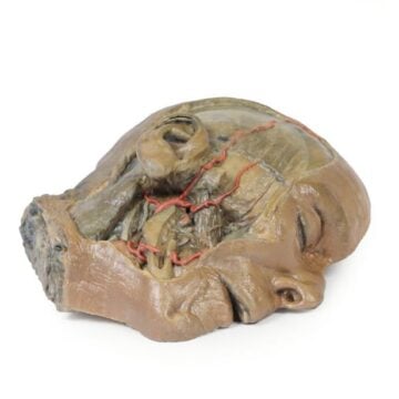
The Superficial Face is a 3D-printed anatomical model crafted for medical educators, students, and clinicians who need clear visualisation of facial neurovascular structures. It solves the challenge of teaching and learning facial anatomy by offering accurate, colour-highlighted modeling of the superficial left face. Choose this model to gain superior clinical accuracy and durable educational quality that enhances anatomical understanding.
Unlock Comprehensive Learning with the Superficial Face 3D Model
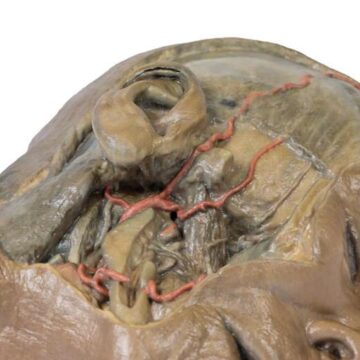
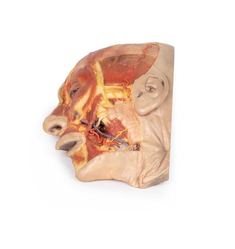
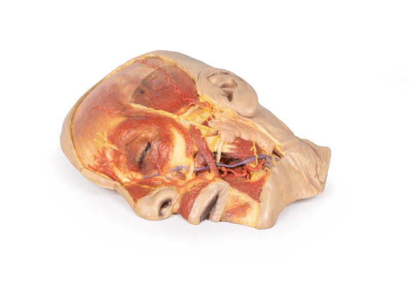
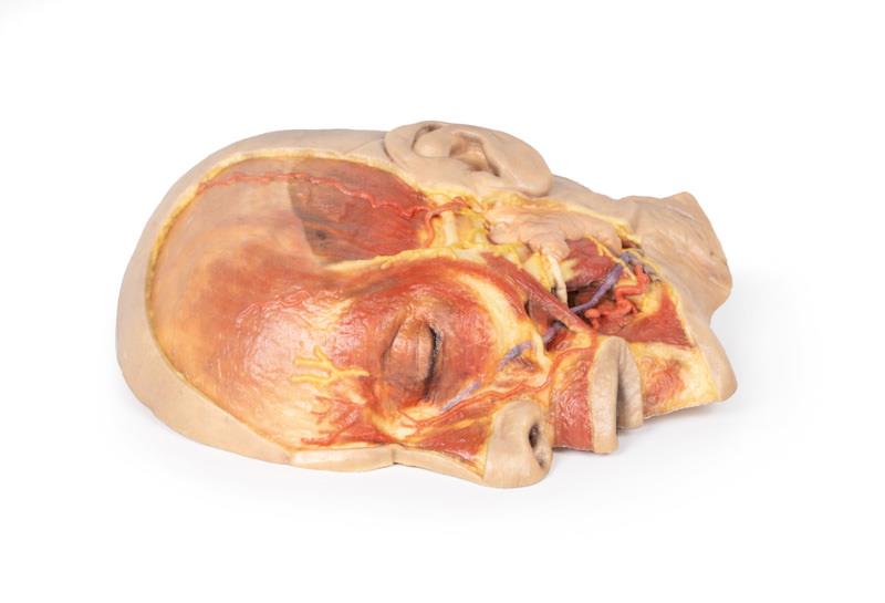
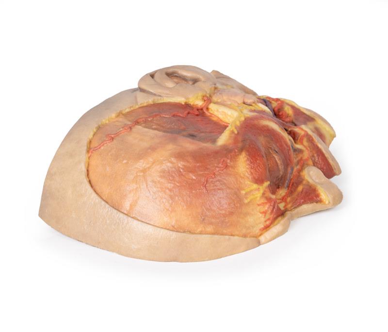
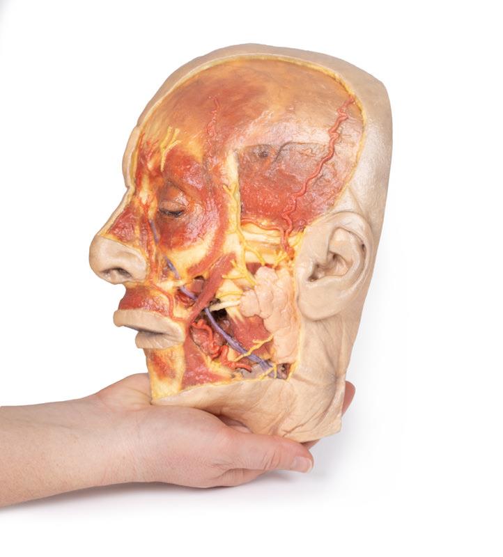
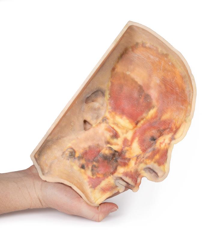
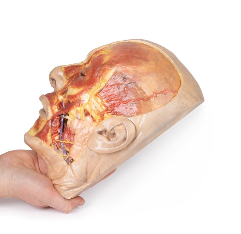
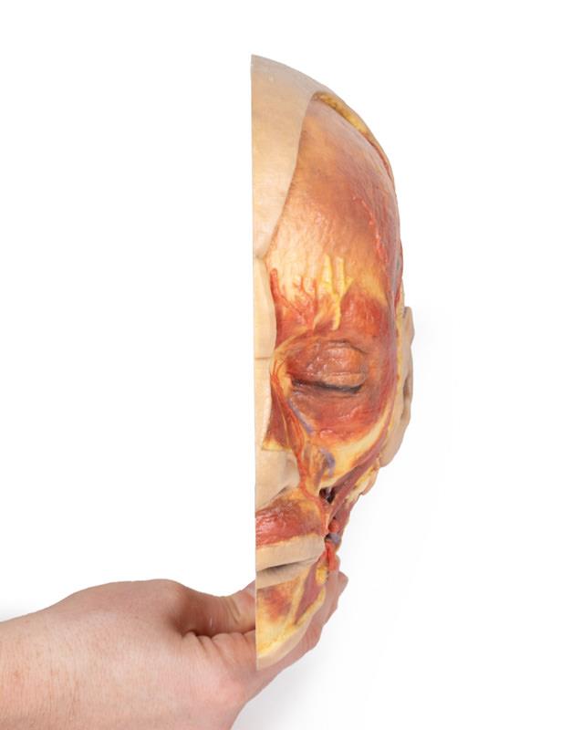
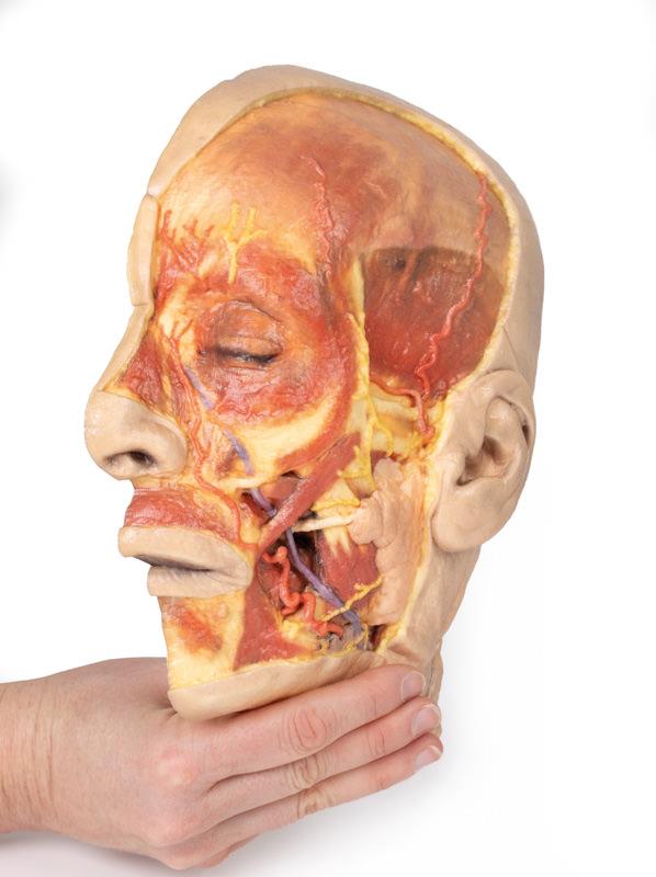
This cutting-edge 3D model captures a meticulous superficial dissection of the left face just anterior to the ear. False colouring highlights critical neurovascular elements and the most important muscles of facial expression. The digitally removed undissected regions provide clear views of the parotid gland, its duct, and the terminal branches of the facial nerve. Users gain unmatched clarity when tracing pathways of structures like the facial artery and vein, or studying the diverse highlighted muscles from the masseter to the orbicularis oculi. Perfect for hands-on anatomy classes, clinical demonstrations, and surgical planning, the Superficial Face model delivers clinical-grade quality for advanced learning.
Features and Benefits
- 3D-printed model with accurate superficial dissection of the left face
- False colouring distinguishes neurovascular structures and superficial facial muscles
- Highlights parotid gland and duct progressing toward the oral cavity
- Displays terminal branches of the facial nerve – cervical, mandibular, buccal, zygomatic, and temporal
- Reveals course of the facial vein and artery from mandible to orbit
- Muscles highlighted include masseter, depressor anguli oris, depressor labii inferioris, zygomaticus major and minor, orbicularis oris, nasalis, levator labii superioris alaeque nasi, procerus, and orbicularis oculi
- Includes the auriculotemporal nerve and superficial temporal artery near the parotid gland
- Shows partially dissected temporal fascia and temporalis muscle
- Highlights supraorbital nerve and supratrochlear and supraorbital arteries on the epicranial aponeurosis
- Features visible layer of the deeper frontalis muscle
- Clinical-grade detail and durable construction for education
Indications for Use
- Anatomy education for students and professionals
- Clinical demonstration of facial nerve and vessels
- Surgical planning and simulation
- Pathway tracing of facial muscles and neurovascular structures
Size Guide
- Model represents left side of adult face, anterior to the ear
- Life-size proportions based on clinical anatomy standards
- Exact SKU – ERLMP1108