

Features
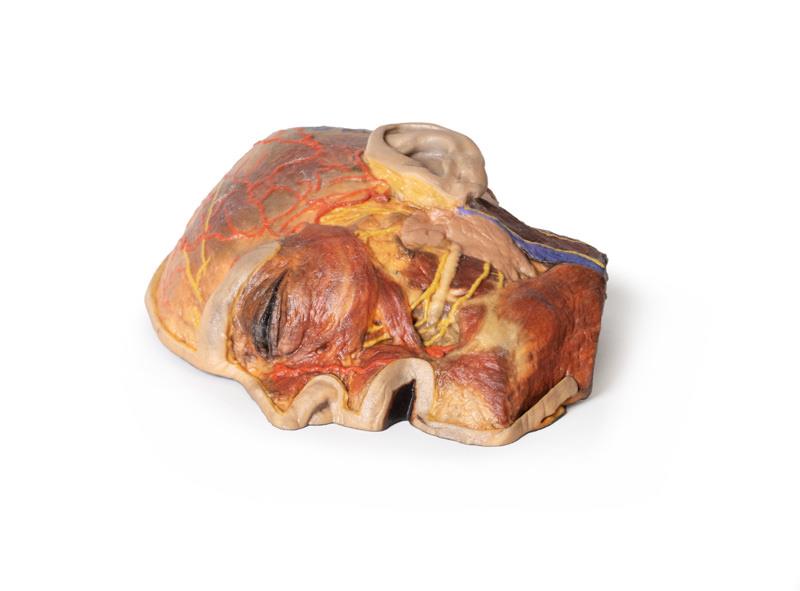
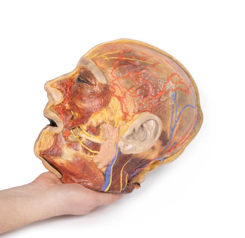
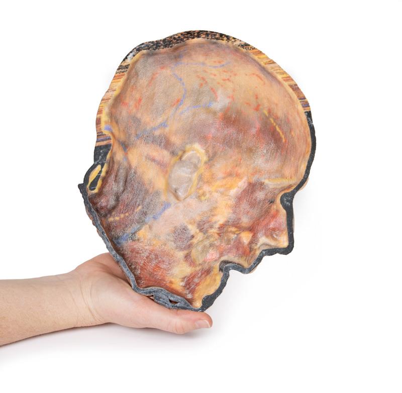
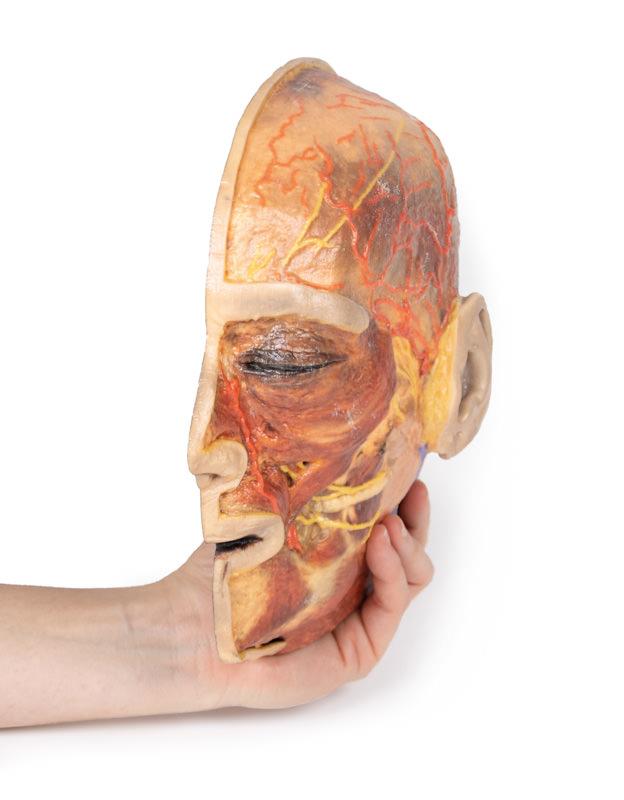
The Superficial Facial nerves & Parotid Gland is a precise 3D printed model crafted for medical educators, clinicians, and students seeking an advanced understanding of facial anatomy. It solves the challenge of visualizing complex superficial neurovascular and muscular structures of the face and head for in-depth learning and effective teaching. Choose this model for its clinical-grade accuracy, expanded anatomical detail, and clear presentation of nerve, artery, vein, and muscle pathways compared to basic models.
Gain Expanded Insight into Facial Anatomy with the Superficial Facial nerves & Parotid Gland Model
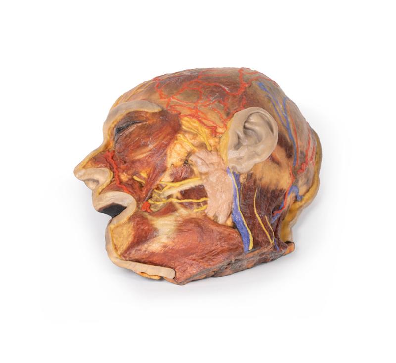
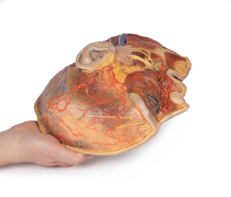
This detailed 3D model elevates facial and head anatomy education by offering an unparalleled dissection of superficial neurovascular and muscular structures. You see an extended course of the facial nerve (CNVII) from the parotid gland and the retained platysma reaching toward the neck. Compared to standard models, this version exposes the anatomy beneath the external ear, the posterior scalp, and occipital region. Educators and students gain superior views of the supraorbital and supratrochlear nerves and the superficial temporal artery, as well as the retromandibular vein and great auricular nerve. This clinical-grade print ensures unmatched reliability for teaching and demonstration.
Features and Benefits
- Expanded dissection of scalp, face, and posterior head
- Clear path of terminal branches of facial nerve (CNVII) from parotid gland
- Retained platysma muscle with extension toward the neck
- Exposed retromandibular vein with visible great auricular nerve fibers
- Detailed display of supraorbital, supratrochlear, lesser occipital nerves, and arteries
- Visible auricularis superior and posterior muscles around ear
- Posterior scalp integration with occipitalis and epicranius muscles
- Clinical-grade 3D print for accurate education and demonstration
Indications for Use
- Anatomy teaching and medical education
- Clinical demonstration for health professionals
- Surgical anatomy training of facial, nerve, and parotid structures
- Medical student learning and exam review
Size Guide
- Model represents life-size adult human facial and head anatomy
- Dimensions match actual anatomical proportions for realistic demonstration