Features
Clinical History
A 36-year old female has a large postpartum hemorrhage after the birth of her 4th child by breech delivery. Her previous 3 children have all been breech deliveries, and no miscarriages. She has a history of intermittent mild abdominal pain. The obstetricians were unable to stop the bleeding and performed an emergency radical hysterectomy and bilateral salpingo-oophorectomy. The patient and baby made a full recovery.
Pathology
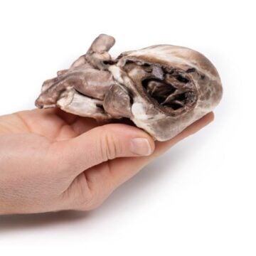
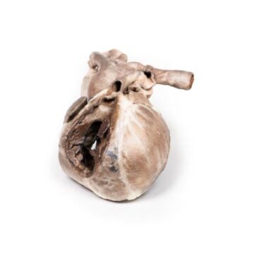
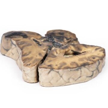
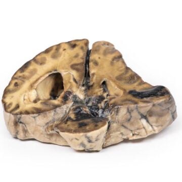
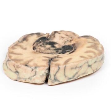
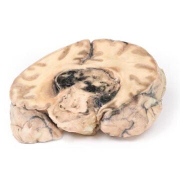
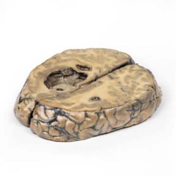
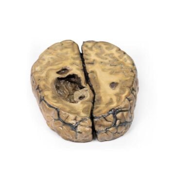
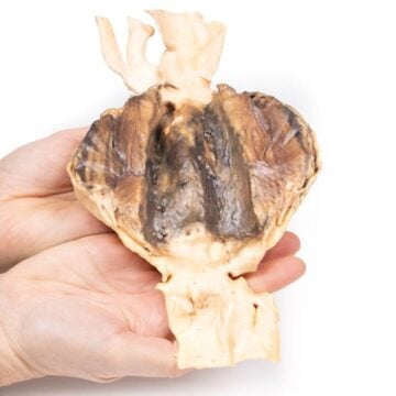
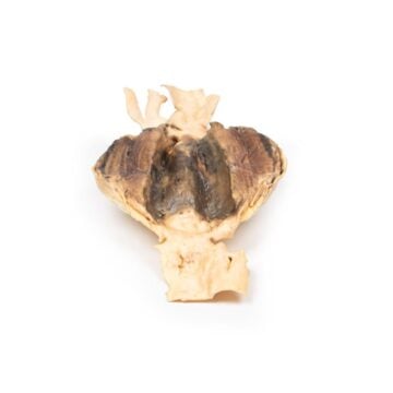
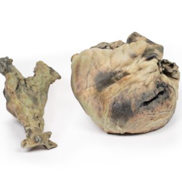
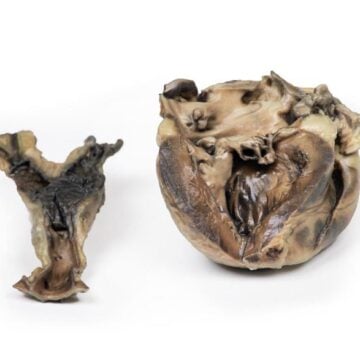
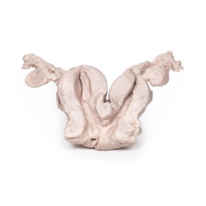
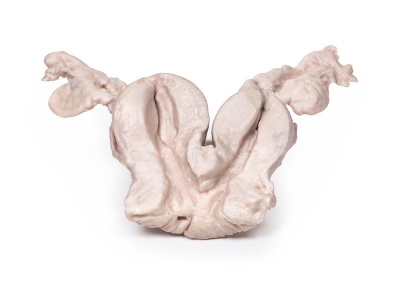
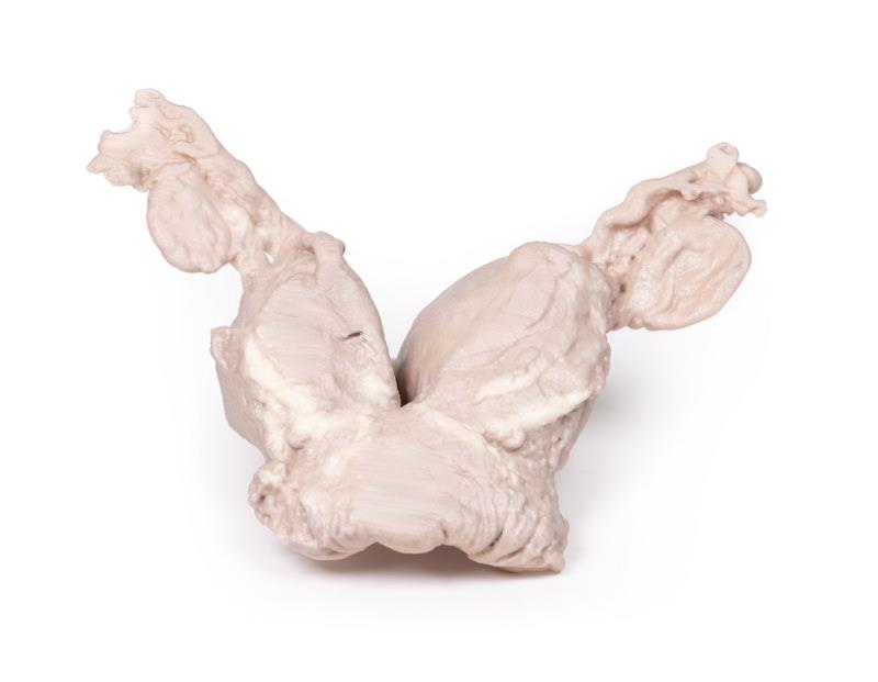
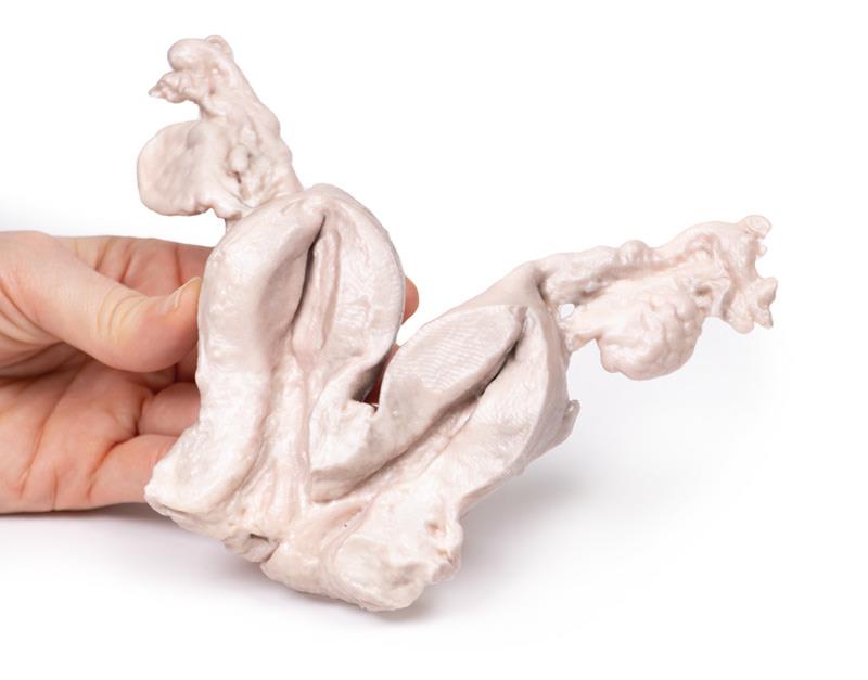
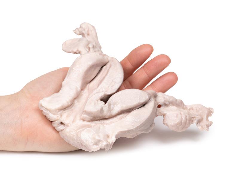
This hysterectomy specimen is of a bicornuate uterus, fallopian tubes and ovaries; sliced coronally and mounted to display cut and external surfaces. Both uterine bodies are equal in size and share a common cervical canal. A few small cysts are present in the cervix.
Further Information
A bicornuate uterus is a congenital uterine malformation where the uterus fundus has an indentation of more than 1 cm. The vagina and cervix are usually normal. There is generally one cervix. There are usually two moderately separate endometrial cavities. Bicornuate uterus develop during embryogenesis when the Mullerian ducts only partially fuse instead of completely fusing.
The risk of developing these malformation increases if exposed to diethylstilbestrol (DHS) in-utero, a synthetic estrogen previously used to prevent pregnancy loss. The karyotype of most women with uterine malformations is 46,XX.
These malformations are present in around 0.5% of women, although the actual number may be greater as not all women are symptomatic. Symptoms may include pelvic pain (cyclic and non-cyclic), abnormal uterine bleeding and discharge and urinary tract infection (UTI). In pregnancy bicornuate uterus may lead to recurrent miscarriage, pre-term labour, fetal growth restriction, fetal malpresentation and placenta previa. Malpresentation of the fetus leads to increased need for caesarean section. There is increased risk postpartum or placental retention and postpartum hemorrhage.
Bicornuate uterus is usually diagnosed with pelvic ultrasound scan. MRI scanning is rarely used to consolidate the diagnosis. Most cases do not require any treatment.