Features
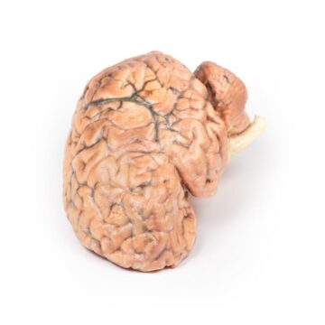
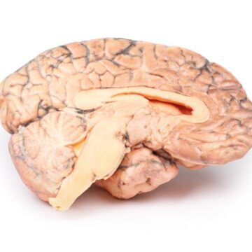
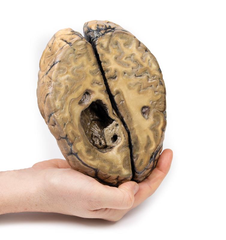
The Metastatic carcinoma in the brain is a premium 3D printed anatomical model crafted for medical educators, trainees, and clinicians. It shows detailed pathological features of brain metastases, enabling an accurate and visual understanding of tumour progression and its neurological impact. This model gives learners a tactile experience, making complex neuro-oncology cases easier to illustrate and comprehend.
Visualise Advanced Tumour Pathology with the Metastatic carcinoma in the brain 3D Model
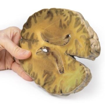
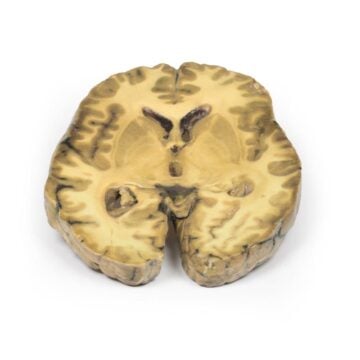
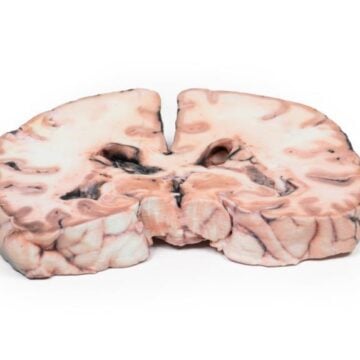
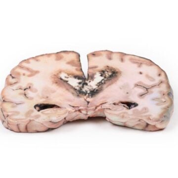
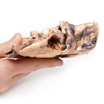
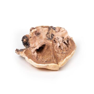
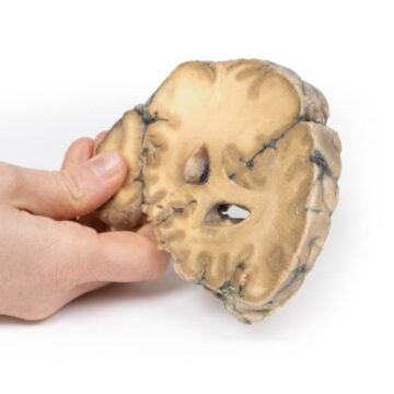
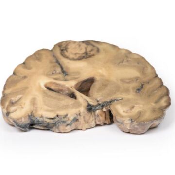
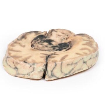
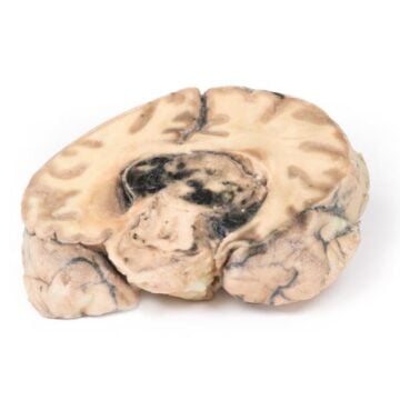
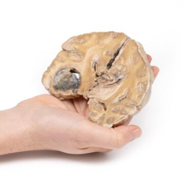
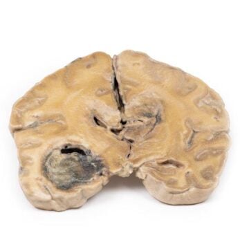
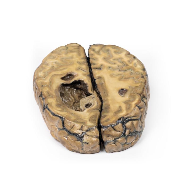
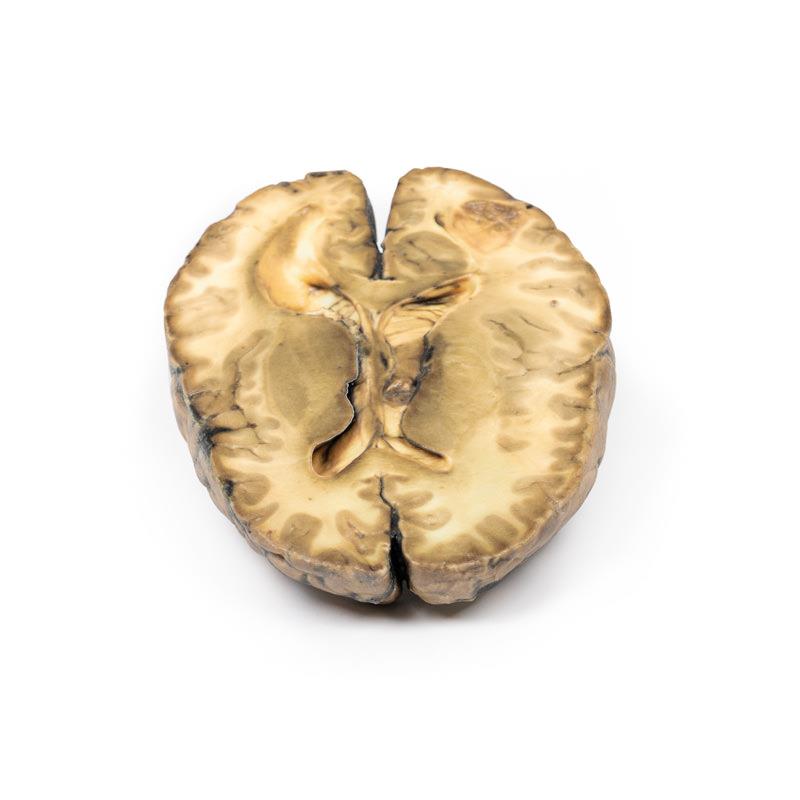
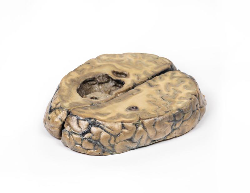
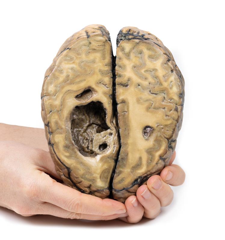
Bring brain metastasis pathology to life with a high-resolution 3D printed model rooted in real clinical history. This product displays authentic horizontal brain slices, clearly revealing space-occupying cystic tumours characteristic of metastatic progression. It features realistic tumour sizes, clear tissue demarcation, and highlights structural changes including ventricular ulceration and brain herniation. Use this model to enhance medical teaching, clinic case discussions, and student examinations. Precision printing and durable materials ensure long-term use in any educational setting.
Features and Benefits
- Authentic cerebrum specimen sliced horizontally for superior anatomical clarity
- Displays three distinct cystic tumours in the parietal region
- Accurately presents tumour sizes – largest 5 cm, secondary 2 x 1.5 cm, third 1.5 cm
- Highlights pathological changes including sub-falcine herniation and ventricular ulceration
- Shows displacement of the basal ganglia and internal capsule
- Manufactured with clinical-grade, durable materials for repeated educational use
- Essential for illustrating metastases from primary breast carcinoma
- Supports high-level medical training and examination demonstrations
Indications for Use
- Ideal for medical and nursing education on brain tumour pathology
- Valuable in neurology and pathology case presentations
- Supports tumour board discussions and interprofessional training
- Assists in anatomical demonstration of metastatic disease progression
Size Guide
- Model based on horizontal slice of human cerebrum
- Largest tumour – 5 cm diameter in right parietal region
- Secondary tumour – 2 x 1.5 cm diameter near posterior margin
- Third tumour – 1.5 cm diameter in left parietal region
- Model SKU – ERLMP2019