Features
The Glioblastoma multiforme is a clinical-grade 3D printed anatomical model designed for healthcare educators, students, and medical professionals. It visualizes the distinct pathological features of one of the most aggressive brain tumors, enabling clear teaching and advanced study. Choose this model for detailed, tactile demonstrations that clarify the complexity of Glioblastoma multiforme, improving comprehension and training outcomes.
Enhance Medical Learning with a Realistic 3D Model
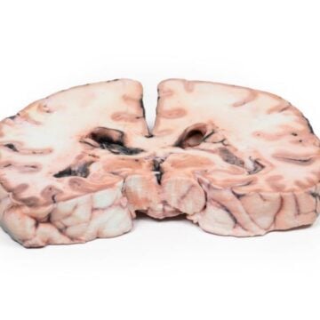
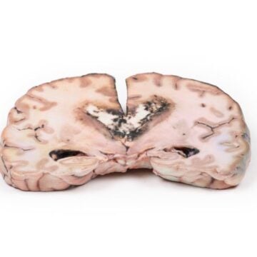
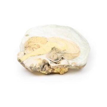
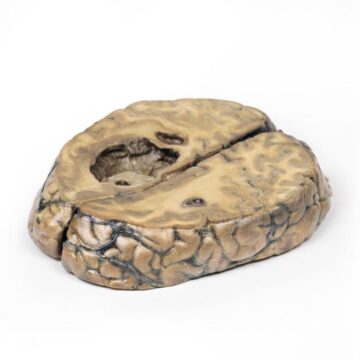
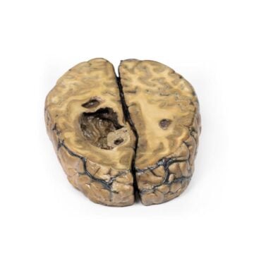
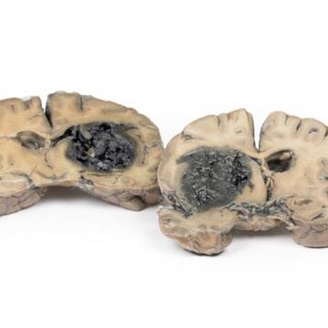
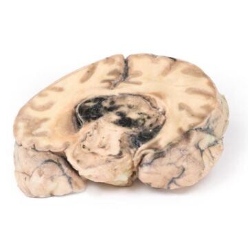
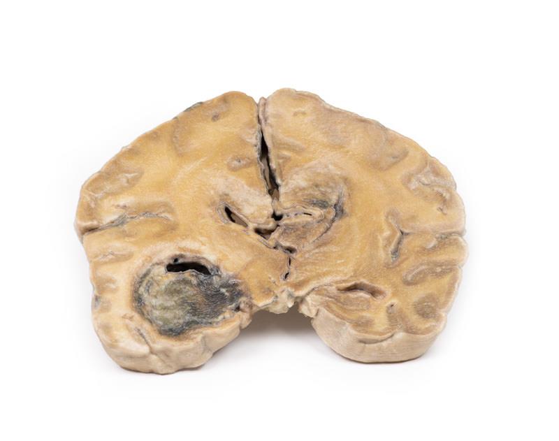
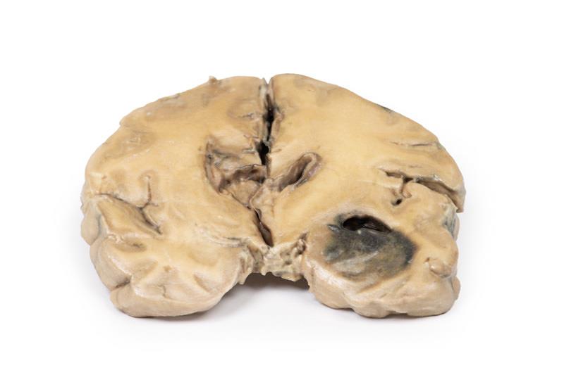
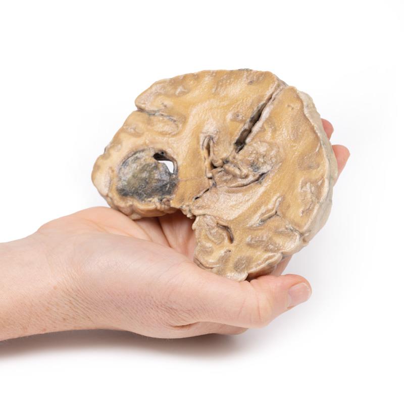
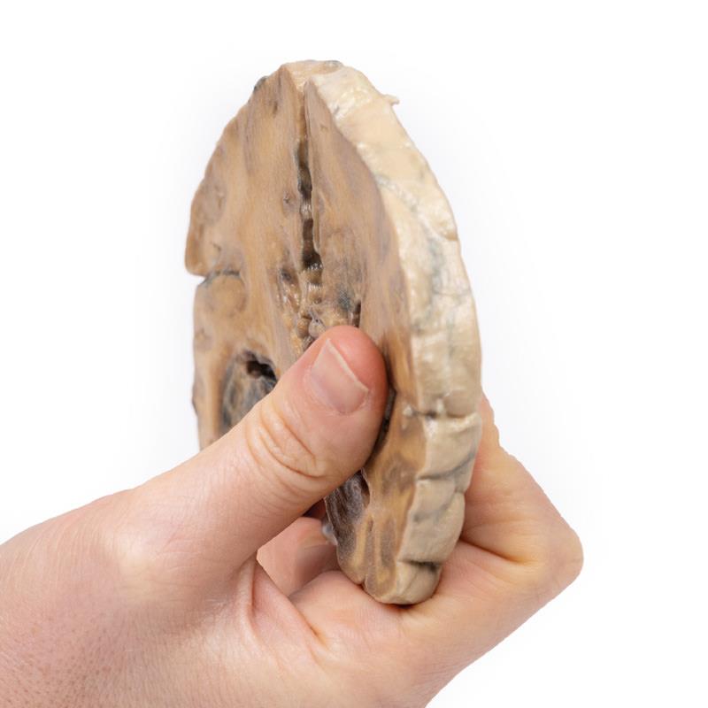
This model captures the unique characteristics of a Glioblastoma multiforme as seen in actual clinical cases. You see clear, variegated tumor masses in the left temporal lobe, including hemorrhagic areas and midline expansion that obliterates the ventricular system. The model illustrates extensive tumor spread across the corpus callosum, reflecting real pathological progression. Clinical-grade detail supports education at the highest level, bridging the gap between textbook descriptions and true anatomical pathology. Durable materials and precision manufacturing ensure that the model remains a reliable teaching aid in any learning environment.
Features and Benefits
- 3D printed from real pathological specimens for maximum accuracy
- Represents hemorrhagic, variegated tumor in the left temporal lobe
- Shows tumor crossing corpus callosum, obliterating ventricles
- High-resolution, clinical-grade model for professional education
- Robust construction for repeated classroom or demonstration use
- Visualizes tumor extensions previously only seen in clinical imaging
Indications for Use
- Medical and allied health student education
- Neuropathology and neuro-oncology training
- Clinical demonstrations to patients and families
- Examination preparation and study
- Professional teaching in lectures and seminars
Size Guide
- Standard model dimensions approximately 16 cm x 12 cm x 10 cm
- Model represents adult human brain proportions and tumor size as observed in pathology
Case Study
Clinical History
Over a 3-year period, a 57-year-old woman had intermittent frontal headache and memory disturbance with progression to psychiatric disturbance, vomiting, and meningeal signs. Localizing neurological signs only developed late in the course of the disease.
Pathology
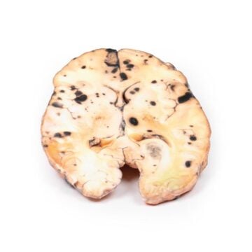
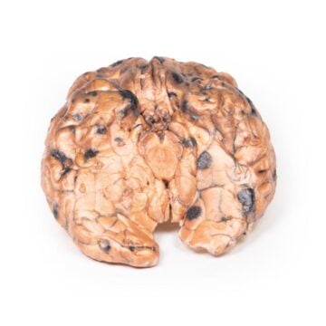
The coronal section through the cerebral hemisphere demonstrates a round, hemorrhagic, variegated tumor in the left temporal lobe. Less well-defined tumor tissue extends across the mid-line replacing the corpus callosum. The ventricular system has been almost totally obliterated. Further sections through the cerebral hemisphere confirmed that these separate lesions are extensions of one massive tumor.
Further Information
Gliomas are the second most common cancer of the central nervous system after meningiomas. The term glioma refers to tumors that are histologically similar to normal glial (macroglia) cells* i.e. astrocytes, oligodendrocytes, and ependymal cells. They arise from a progenitor cell that differentiates down one of the cell lines. GBMs can arise in the brain de novo or evolve from lower grade astrocytoma or oligodendrogliomas. GBM is often referred to as grade IV astrocytoma. They are differentiated histologically from anaplastic astrocytoma by necrotizing tissue surrounded by anaplastic cells as well as by the presence of hyperplastic blood vessels.
GBMs are more common in males. It is most diagnosed in the sixth decade of life. Genetic risk factors include neurofibromatosis type 1 and Li-Fraumeni syndrome. Previous brain radiotherapy is also associated with increased risk of GBM. Symptoms vary depending on the location of the GBM, but may include any of the following:
Persistent headaches
Double or blurred vision
Vomiting
Loss of appetite
Changes in mood and personality
Changes in ability to think and learn
New onset of seizures
Speech difficulty of gradual onset
Diagnostic tools include computed tomography (CT scan) and magnetic resonance imaging (MRI). Around 50% of these tumors occupy more than one cerebral hemisphere. GBMs commonly extend into the ventricular walls or meninges, and thus into the central spinal fluid (CSF). Spinal cord spread is uncommon. Metastasis beyond the central nervous system is rare. Tumor growth causes cerebral oedema leading to increased intra-cranial pressure. These are biologically aggressive tumors, and if left untreated survival is typically 3 months. The mainstay of treatment for GBMs is surgery, followed by radiation and chemotherapy.
*Microglia are a different lineage from macroglia. The former is related to the macrophage lineage and arise initially from the yolk sac and later in development from the bone marrow.