
Features
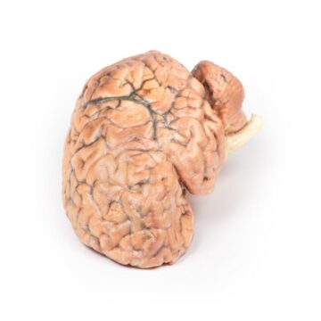
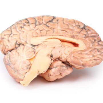
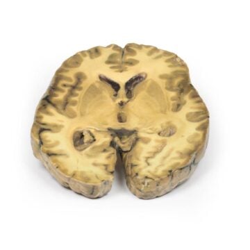
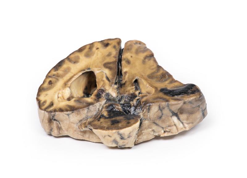
The Intracranial space-occupying lesion is a high-fidelity 3D printed anatomical model designed for medical professionals, educators, and students. It illustrates the brain’s structural changes caused by a right-sided expanding lesion, such as a meningioma. This tool helps target advanced neuroanatomy teaching by providing a tangible, clinically accurate representation of critical pathological features that improve understanding of neurological deficits and herniation syndromes.
Enhance Learning with a Realistic Brain Pathology Model
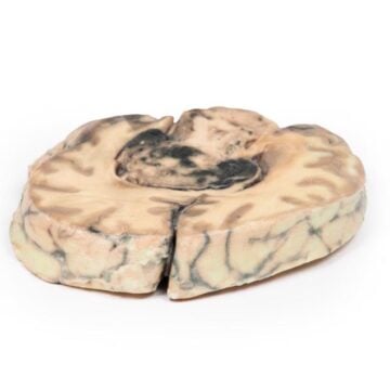
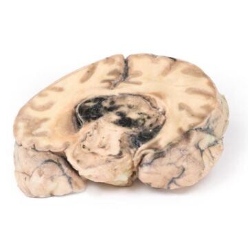
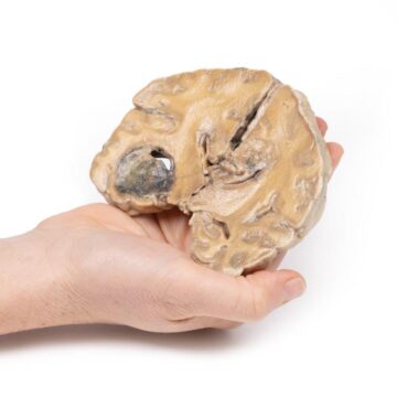
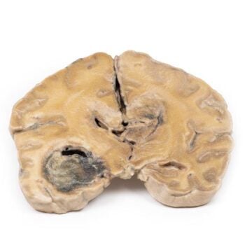
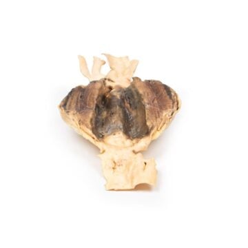
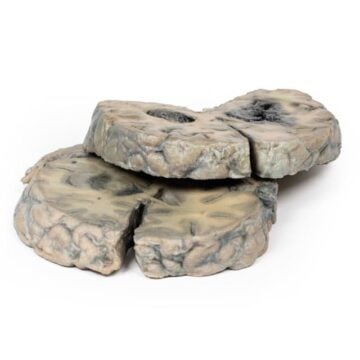
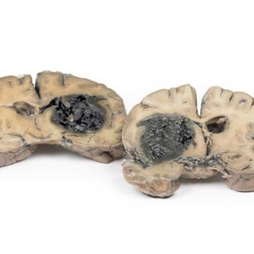
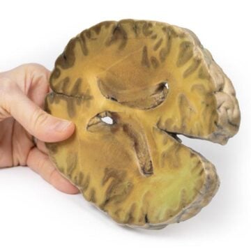
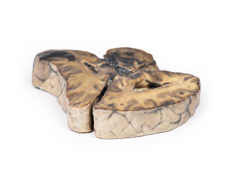
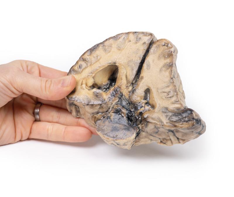
This detailed Intracranial space-occupying lesion model demonstrates the brain’s response to a mass effect, including midline shift, subfalcine herniation, and varying stages of hemorrhage. The clinically accurate replica captures features seen in a patient with repeated headaches, seizures, and eventual coma, making the abstract clinical history visible and tangible. Educators and learners benefit from seeing ventricular asymmetry, displaced cingulate gyrus, and temporal lobe hemorrhage, reinforcing theory with precise anatomical visuals. Designed to meet clinical-grade and educational standards, the model helps users correlate neuroanatomical changes with patient symptoms and improves retention of complex pathology concepts.
Features and Benefits
- Accurate 3D printed coronal brain section featuring real patient pathology
- Displays midline shift and subfalcine herniation of the cingulate gyrus
- Highlights hemorrhage of varying ages in the temporal lobe and pons
- Shows ventricular asymmetry and brain parenchyma displacement
- Clinical-grade quality for education and demonstration
- Ideal for visualizing mass effect and herniation syndromes
- Constructed for durability in repeated teaching scenarios
Indications for Use
- Medical and health science education
- Demonstrations of brain mass effects and herniation patterns
- Neurology, pathology, and neurosurgery teaching
- Patient and student education on intracranial lesions
Size Guide
- Full-scale coronal brain section based on human anatomical dimensions
- Width – standard adult brain section (approximately 13-15 cm)
- No alternate sizes or ranges—model matches typical human adult brain anatomy