Features
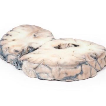
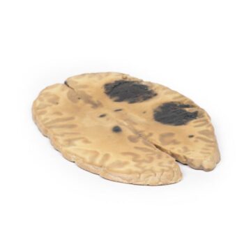
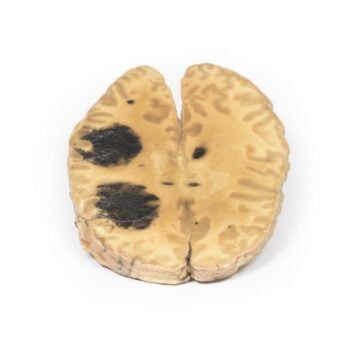
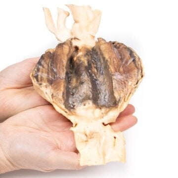
The Berry Aneurism of Basilar Artery is a precision 3D anatomical model designed for medical educators, clinicians, and students. It presents detailed pathology essential for understanding aneurysm complications and surgical challenges. This model serves as an exceptional educational resource by illustrating a real patient case with remarkable anatomical accuracy.
Enhance Clinical Training with the Berry Aneurism of Basilar Artery 3D Model
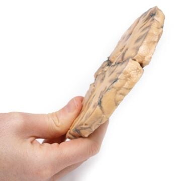
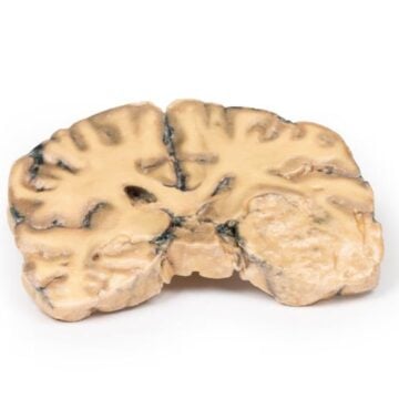
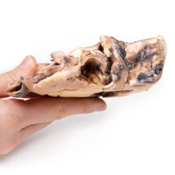
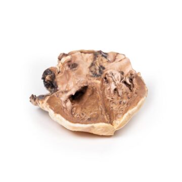
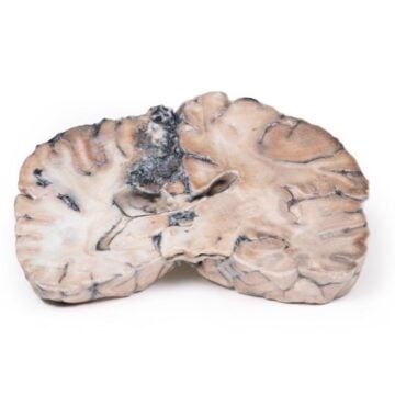
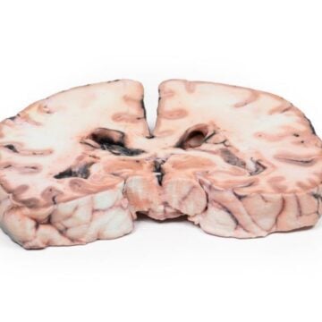
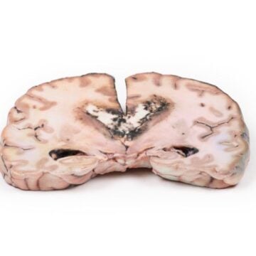
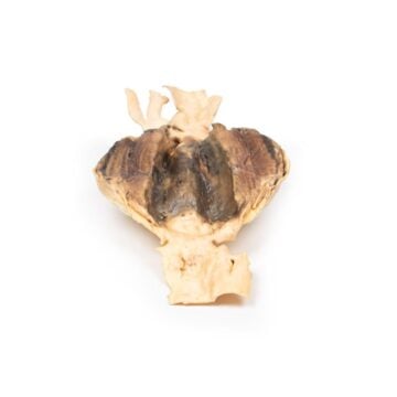
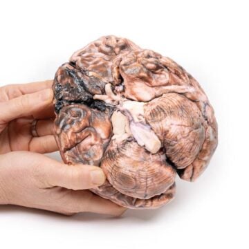
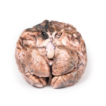
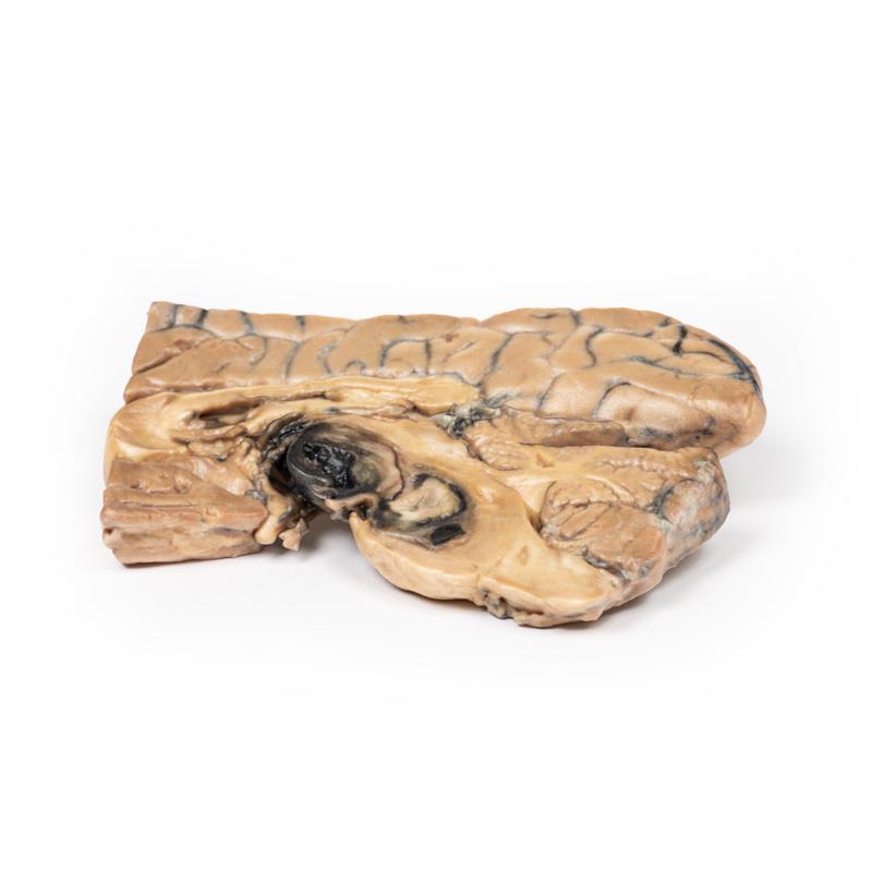
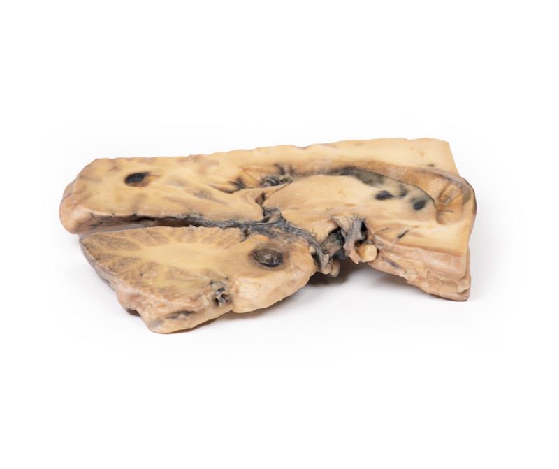
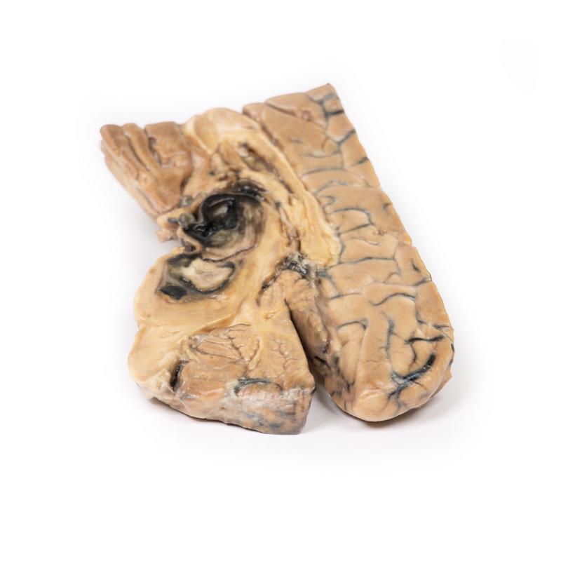
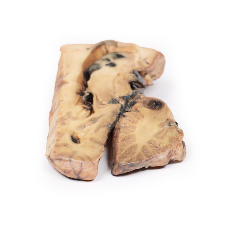
This model showcases a life-sized, mid-sagittal brain hemi-section with a large, darkly-colored ovoid aneurysm arising from the basilar artery. Medical professionals use this model to study rare vascular anomalies, improve diagnostic skills, and simulate intervention strategies for complex neurovascular cases. Every visible anatomical feature, including a laminated thrombus and mass effect on adjacent brain structures, helps users understand the far-reaching clinical impact of basilar artery aneurysms. Choose this model for its fidelity, educational value, and direct relevance to both training and patient education.
Features and Benefits
- Highly detailed 3D-printed mid-sagittal brain slice showing a 5 x 2 cm berry aneurysm
- Displays laminated thrombus within the aneurysm
- Accurately depicts mass effect on the midbrain, third ventricle, and pons
- Includes area of mucoid degeneration behind the aneurysm
- Shows dilated lateral ventricles and patchy haemorrhagic infarction of the caudate nucleus
- Highlights blood clot in the third ventricle and signs of subarachnoid haemorrhage
- Clinical-grade print ideal for hands-on teaching and demonstration
- Valuable for case-based learning and neurosurgical simulation
Indications for Use
- Medical and allied health education
- Clinical anatomy teaching for neurovascular conditions
- Student and resident case-based learning
- Simulation of neurosurgical procedures
- Patient education regarding aneurysm complications
Size Guide
- Model thickness – approximately 1 cm (whole brain hemi-section)
- Aneurysm size – 5 x 2 cm diameter
- Mucoid degeneration area – 0.4 cm diameter within pons
- Displays structures including third ventricle, midbrain, pons, lateral ventricles, caudate nucleus