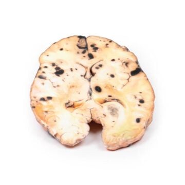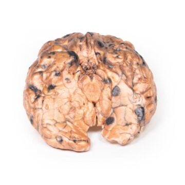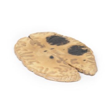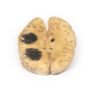
Features
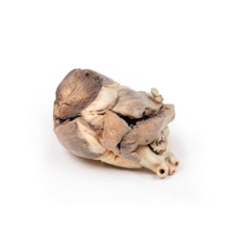
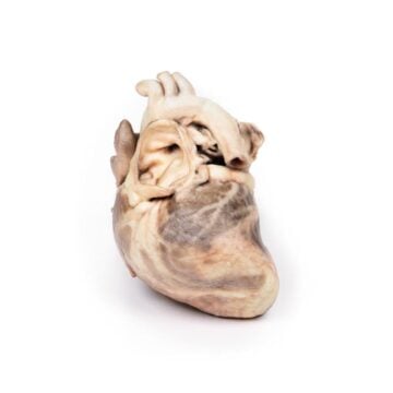
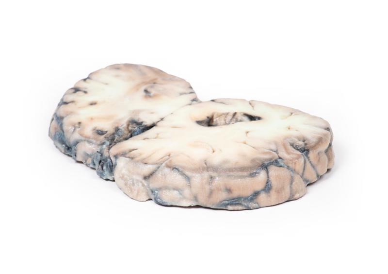
The Cerebral Arterio-Venous Malformation is a clinical-grade 3D printed anatomical model crafted for medical educators, students, and healthcare professionals. It offers a true-to-life replica of a complex brain lesion for in-depth anatomical study and demonstration. This model helps users to better understand and explain pathological brain structures, supporting precise education and improved surgical planning.
Enhance Neuroanatomy Teaching with the Cerebral Arterio-Venous Malformation 3D Model
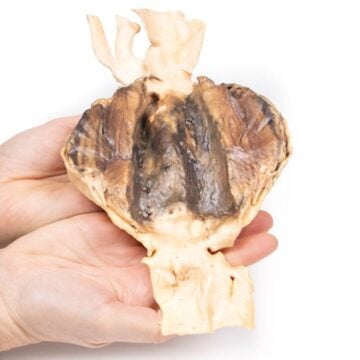
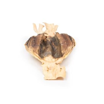
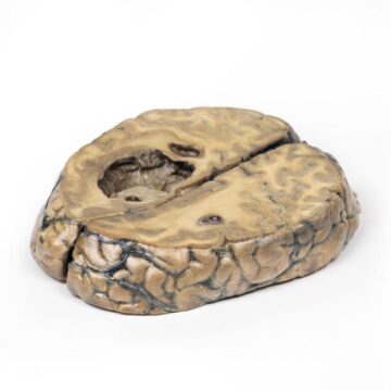
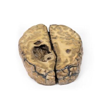
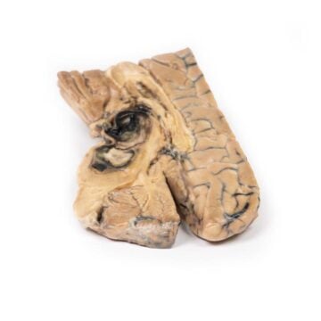
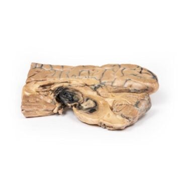
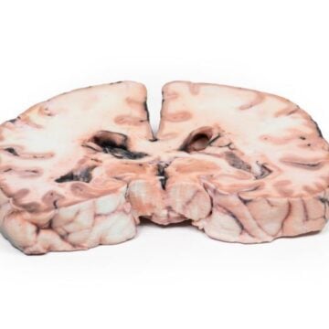
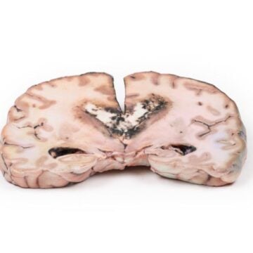
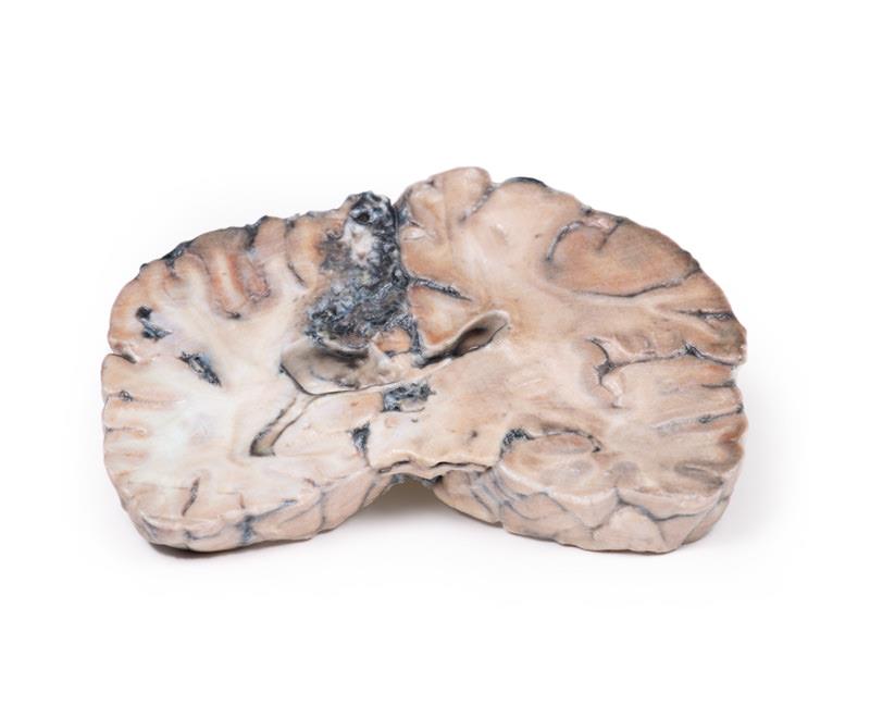
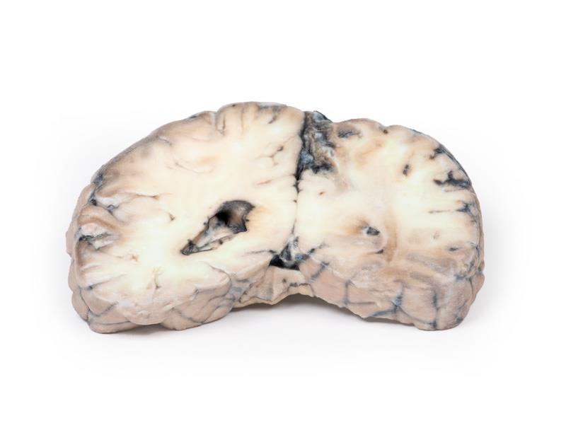
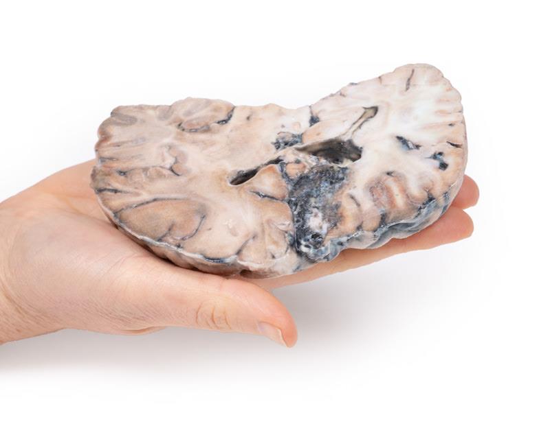
Bring hands-on realism to your classroom or clinical training sessions with this expertly designed 3D printed model. The Cerebral Arterio-Venous Malformation recreates an actual coronal brain section, showing the characteristic tangled vascular channels and abnormal tissue replacement found in AVMs. This physical model reveals pathological changes, such as glial tissue surrounding dilated arteries and veins, vital for teaching diagnosis, symptoms, and possible complications like intracranial hemorrhage or neurological deficit. Its clinical history and accurate scale support advanced understanding of AVM pathology and associated neurological outcomes.
Features and Benefits
- Precise 3D printed coronal brain slice through parietal lobes
- Displays a 4 cm mass replacing cortex and white matter
- Detailed network of tortuous vascular channels visualized
- Shows both arteries with thick walls and veins with thin walls
- Clinical-grade accuracy for advanced education and training
- Suitable for demonstration, patient education, and surgical planning
Indications for Use
- Medical and neuroanatomical education
- Demonstration of cerebral arteriovenous malformations
- Training and planning for surgical and clinical procedures involving AVMs
- Patient communication for AVM symptoms and risks
- Study of AVM-associated complications – headaches, seizures, neurological deficits, and hemorrhage
Size Guide
- Model is a coronal brain slice with a lesion 4 cm in greatest diameter
- Reproduces the actual anatomical dimensions from the specimen, including lesion spanning superior surface to lateral ventricle