Features
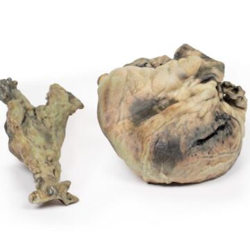
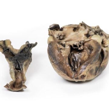
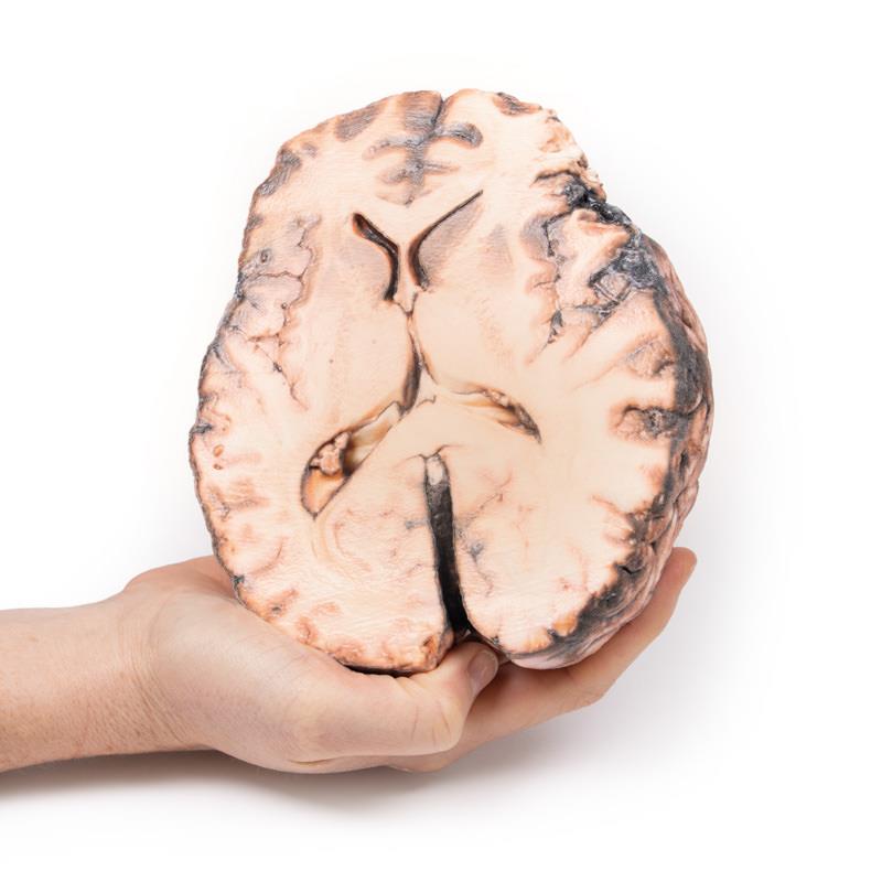
The Ruptured Berry Aneurysm is a clinical-grade 3D printed anatomical model designed for medical professionals, educators, and students who require precise neurovascular pathology representation. It demonstrates the complex presentation and consequences of a ruptured intracerebral aneurysm, supporting advanced education and hands-on training. This model provides unparalleled detail that enhances the teaching of neurological events and surgical interventions.
Enhance Neurovascular Education with the Ruptured Berry Aneurysm Model
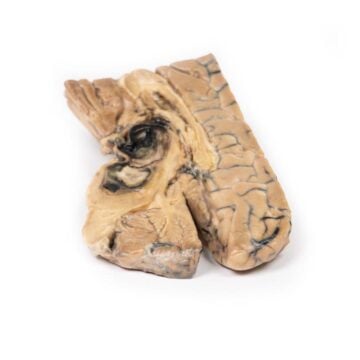
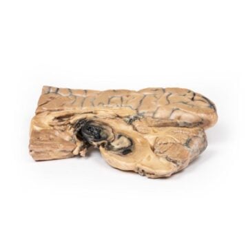
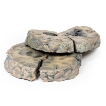
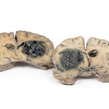
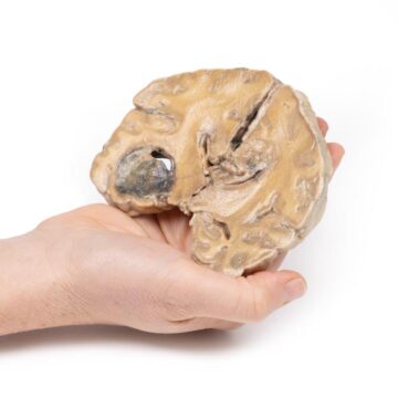
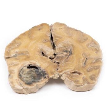
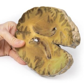
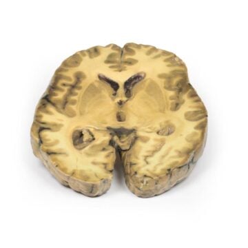
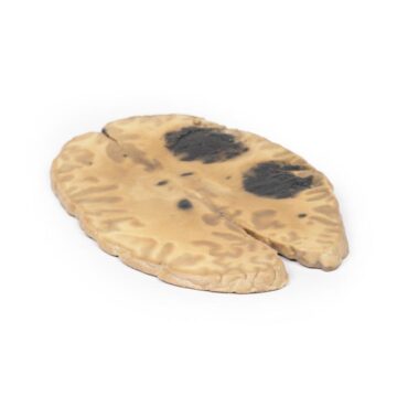
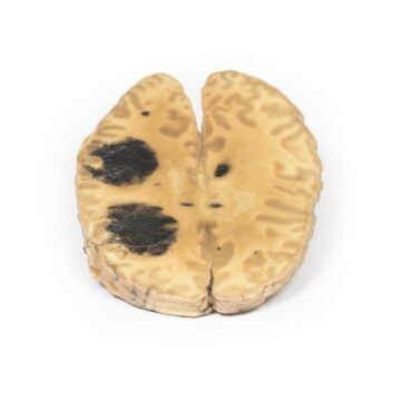
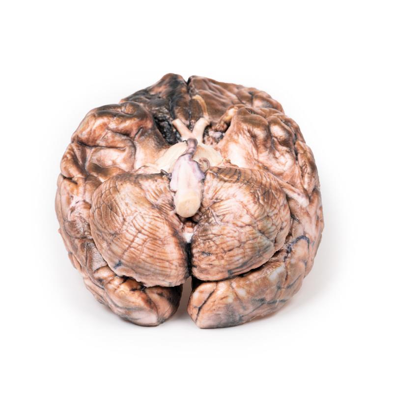
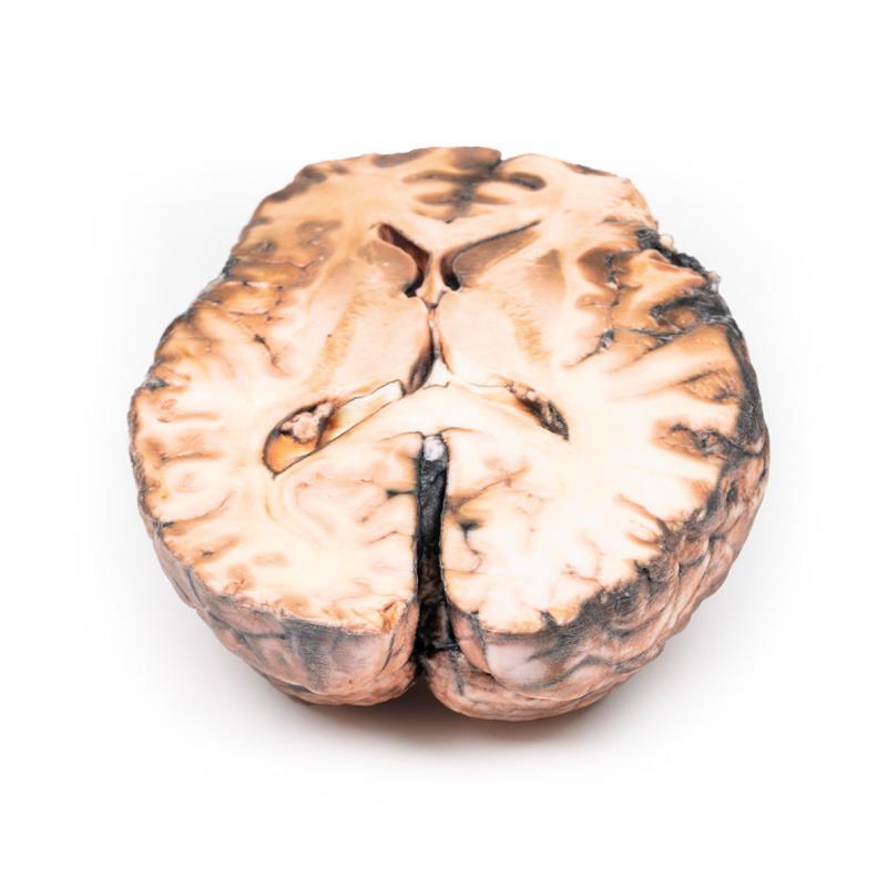
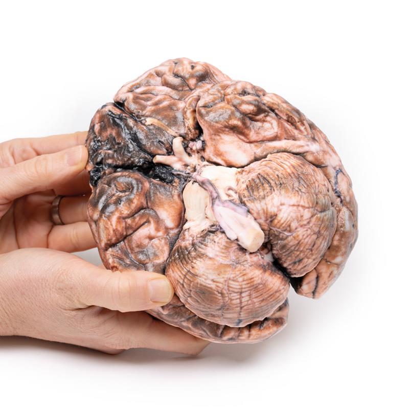
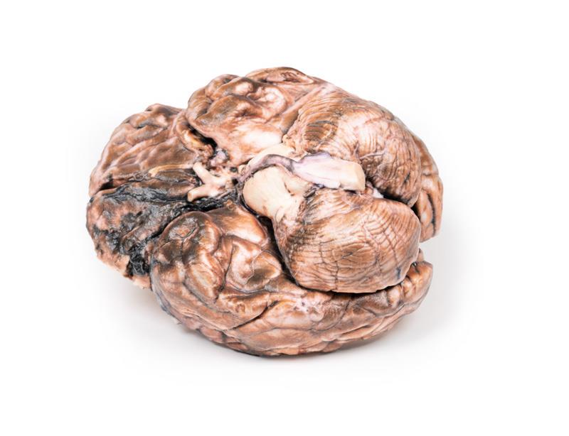
Experience unmatched anatomical accuracy with the Ruptured Berry Aneurysm. This model showcases a saccular aneurysm at the junction of the right internal carotid and posterior communicating artery. The construction replicates a 5 mm wide rupture, with clear display of subarachnoid blood distribution in the cisterna magna and on the right frontal lobe’s inferior surface. It enables users to visualize associated neurological findings such as cranial nerve involvement, frontal lobe softening, and the presence of an unruptured aneurysm on the left side. The model serves as an invaluable tool for real-world scenario simulation, enhancing understanding of clinical pathologies and the neurological impact of vascular defects.
Features and Benefits
- Accurately depicts a saccular aneurysm ruptured at the right internal carotid and posterior communicating artery junction
- Shows subarachnoid hemorrhage in the cisterna magna and right frontal lobe
- Presents dual pathology with an additional unruptured left-sided aneurysm
- Exhibits frontal lobe softening and friability due to compromised blood supply
- Offers clinical-grade, education-standard 3D printing for reliable anatomical study
- Ideal for demonstration of complex neurovascular events, nerve palsies, and vascular-induced neurological deficits
Indications for Use
- Medical and anatomical education related to intracerebral aneurysms
- Clinical training for neurosurgical planning and stroke assessment
- Simulation of cranial nerve pathologies and visual field defects
- Patient education for understanding neurovascular conditions
Size Guide
- Model displays a 5 mm diameter aneurysm at the right internal carotid and posterior communicating artery junction
- Represents a second, unruptured aneurysm on the left side
- Full brain basal surface with pathological and anatomical detail to scale