


Features
Product Description
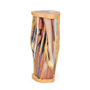
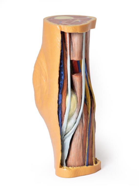
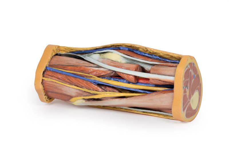
This 3D printed Popliteal Fossa preserves the distal thigh and proximal leg, dissected posteriorly to demonstrate the contents of the popliteal fossa and surrounding region. The proximal cross-section demonstrates the anterior, posterior and medial compartment muscles, with the femoral artery and vein visible within the adductor canal. The sciatic nerve and great saphenous vein are also visible.
The skin, superficial fascia, and deep fascia have been removed over the popliteal fossa to expose the contents of the space. The muscular borders of the space are intact except for a window cut into the semimembranosus muscle to allow a view of the popliteal artery and vein near the adductor magnus. On the medial aspect of the window the great saphenous vein descends on the surface of the sartorius muscle. Distally the sartorius is visible joining the semitendinosus and semimembranosus muscles to form the pes anserinus. All major deep and superficial nerves and vessels of the space are visible, including the superior lateral genicular artery passing towards the anterior compartment of the thigh. Along the lateral margin the posterior aspect of iliotibial tract is visible descending to the lateral epicondyle of the tibia.
The distal cross-section demonstrates the continuation of popliteal contents and branches. The great saphenous and small saphenous veins are visible within the superficial fascia, as are the medial and lateral sural cutaneous nerves. Between the muscles of the posterior, lateral, and anterior compartments are the neurovascular bundles of the leg (posterior tibial artery, veins and tibial nerve; peroneal artery and veins; anterior tibial artery, veins and deep peroneal nerve; superficial peroneal nerve).
This Popliteal Fossa model is 5x4x10 inches. The circumference measuring around the patella is 12 inches.
Made to Order
Please Note: Thanks to the flexibility of manufacturing that 3D Printing offers, this model is “”printed to order””, and is not typically available for immediate shipment. Most models are printed within 15 working days and arrive within 3-5 weeks of ordering, and once an order is submitted to us, it cannot be canceled or altered. Please contact us if you have specific a specific delivery date requirement, and we will do our best to deliver the model by your target date.
Advantages of 3D Printed Anatomical Models
- 3D printed anatomical models are the most anatomically accurate examples of human anatomy because they are based on real human specimens.
- Avoid the ethical complications and complex handling, storage, and documentation requirements with 3D printed models when compared to human cadaveric specimens.
- 3D printed anatomy models are far less expensive than real human cadaveric specimens.
- Reproducibility and consistency allow for standardization of education and faster availability of models when you need them.
- Customization options are available for specific applications or educational needs. Enlargement, highlighting of specific anatomical structures, cutaway views, and more are just some of the customizations available.
View the full range of 3D Printed Anatomical Models here