
Features
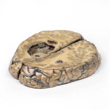
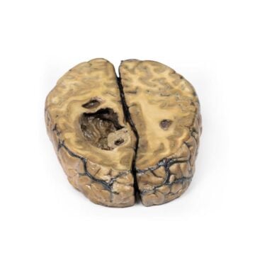
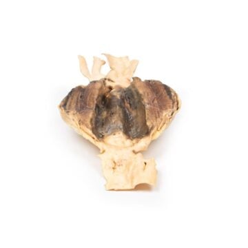
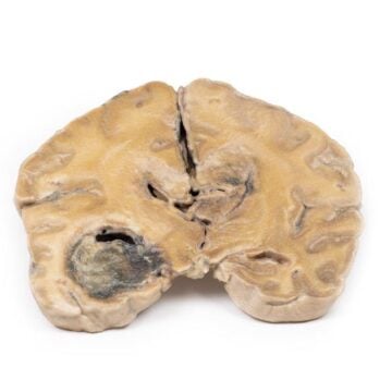
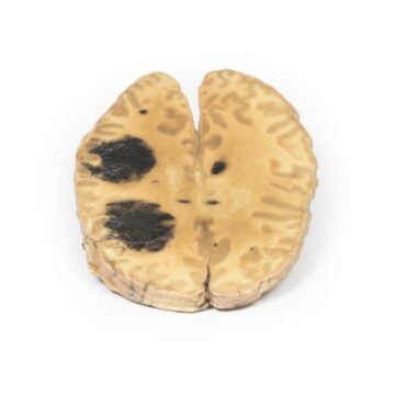
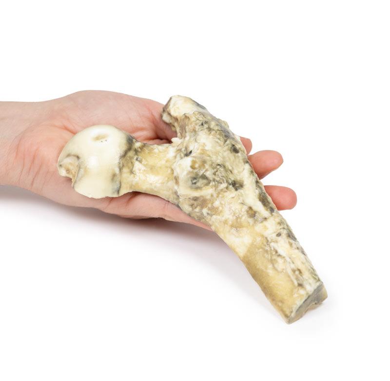
The Metastatic Malignant Melanoma is a clinical-grade 3D printed anatomical model designed for medical educators and healthcare professionals. It accurately depicts metastatic melanoma involving the femur, giving a detailed, realistic view to aid in teaching cancer pathology and metastatic patterns. Choose this model to provide your students or team with a hands-on, visual experience that illustrates the severe impact of melanoma on bone structure and tissue integrity.
Visualize Advanced Melanoma Pathology with Metastatic Malignant Melanoma 3D Model
This 3D printed model brings cancer pathology to life, representing a longitudinally-sectioned human femur infiltrated with metastatic melanoma deposits. It displays the destruction of cancellous bone, the spread of tumor through the medullary cavity, and distinctive pigment variations that range from pale brown to black. Educators and clinicians use it to demonstrate common features of melanoma spread, support cancer awareness, and improve the understanding of metastatic mechanisms. With clinical background accuracy, this model illustrates why early detection and staging remain critical in melanoma care.
Features and Benefits
- Life-size, high-fidelity 3D printed anatomical model based on real patient pathology
- Shows a femur with medullary cavity filled by metastatic melanoma of varying colors up to 3 cm diameter
- Depicts complete destruction of cancellous bone by tumor masses
- Demonstrates thickened, discolored cortical bone at the shaft-neck junction
- Displays diffuse marrow infiltration with clear demarcation between affected and unaffected bone
- Durable and made for repeated educational or clinical demonstration
- Suited for universities, teaching hospitals, clinics, and student study
- Enhances the ability to explain diagnostic, pathological, and therapeutic concepts visually
Indications for Use
- Medical and allied health education
- Pathology demonstrations and teaching tumor spread
- Clinician-patient explanations of metastatic disease
- Revision and study for anatomy and pathology exams
- Research presentation and anatomical display
Size Guide
- Life-size anatomical replication of a proximal right femur
- Tumor deposits up to 3 cm maximum diameter visible in medullary cavity
- Medullary cavity and cancellous bone replicates pathological destruction as seen in advanced metastatic cases