


Features
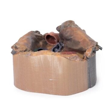
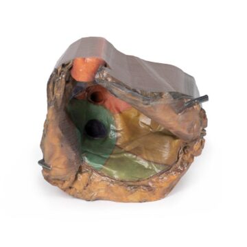
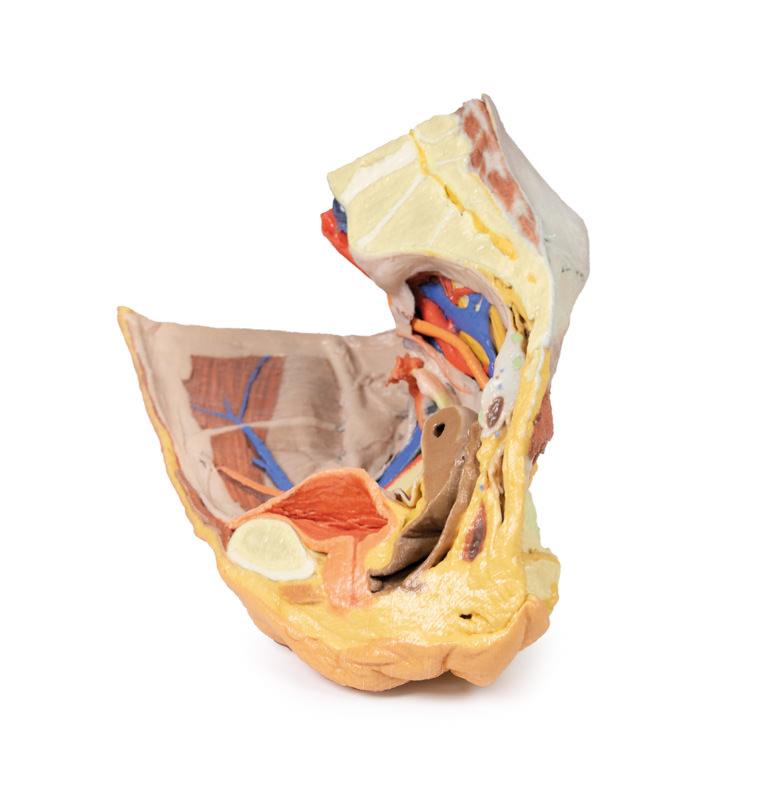
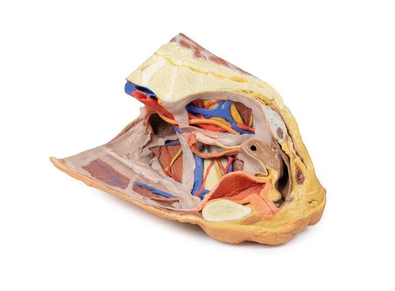
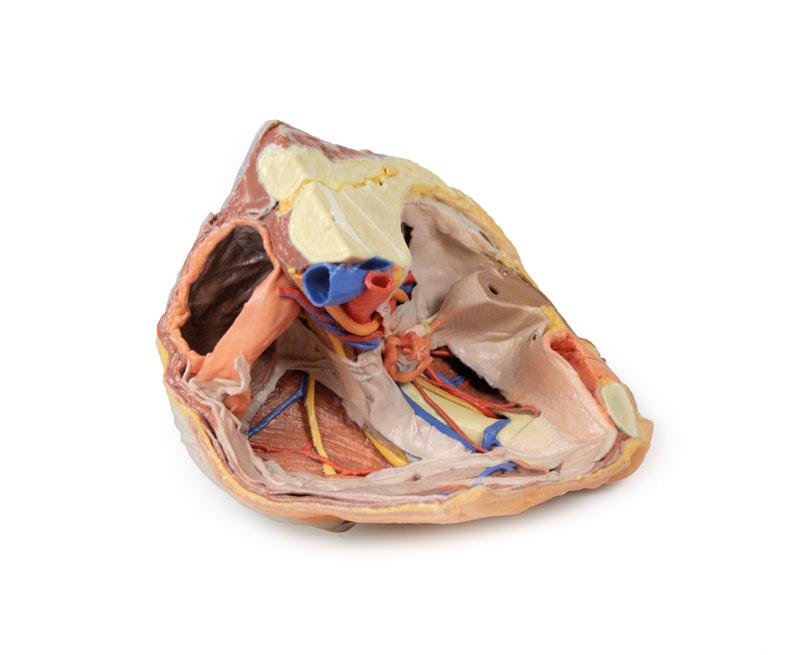
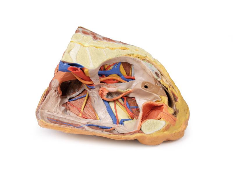
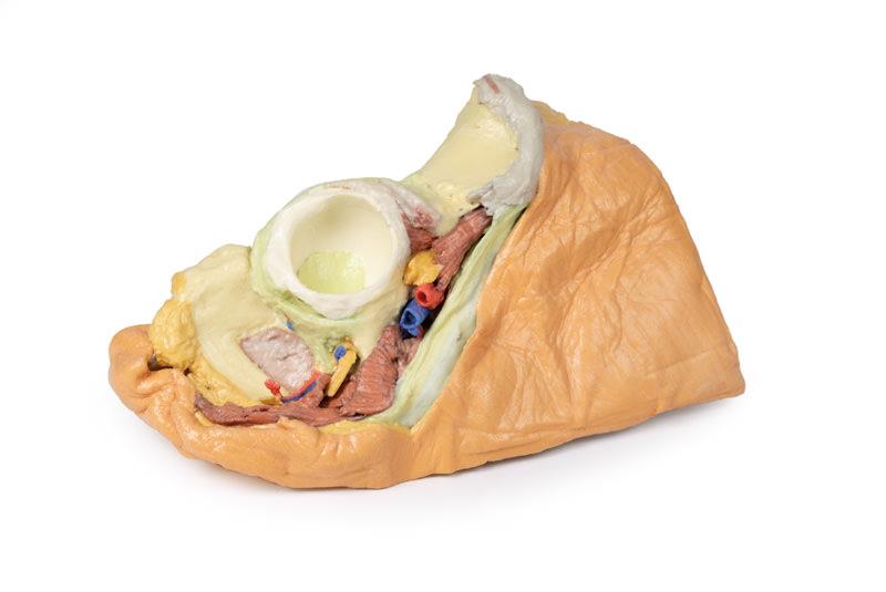
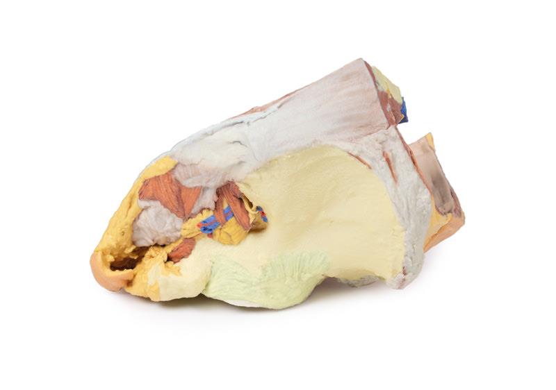
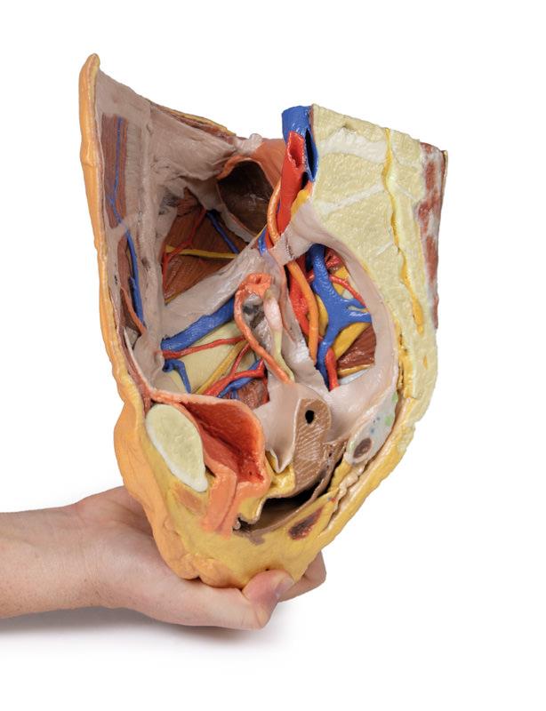
The Female right pelvis superficial and deep structures is a detailed 3D printed anatomical model designed for clinicians, educators, and students seeking in-depth pelvic anatomy visualization. It solves the challenge of demonstrating both superficial and deep aspects of the female pelvis in one accurate replica. This model ensures clinical-grade quality and unmatched detail, enabling clear instruction and study unavailable in basic skeleton models.
Enhanced Realism and Precision for Advanced Pelvic Anatomy Education
Gain unparalleled access to true and false pelvis structures with the Female right pelvis superficial and deep structures model. High-fidelity 3D printing preserves intricate bone, ligament, and membrane anatomy with clinical accuracy. Partial peritoneum removal creates visual ‘windows’, making it easy to identify extraperitoneal structures that standard models often obscure. This unique approach gives educators and learners clear views for explanation and practice. Built for rigorous use, this model withstands frequent handling in demanding institutional settings. It sets a new bar for realism, setting your laboratory or classroom apart.
Features and Benefits
- 3D printed with clinical-grade precision for education-standard anatomy
- Displays both superficial and deep aspects of true and false pelves
- Shows the inguinal ligament, obturator membrane and canal
- Includes greater and lesser sciatic foramina
- Features visual ‘windows’ by removing sections of peritoneum for extraperitoneal views
- Durable for frequent, hands-on use in clinics and classrooms
Indications for Use
- Anatomy education in medical and allied health programs
- Surgical training requiring pelvic structure identification
- Patient education for pelvic surgeries
- Demonstration in academic and research settings
Size Guide
- Life-size model representing adult female right pelvis
- Accurately depicts true and false pelvic boundaries
- Includes superficial and deep anatomical features at 1 -1 scale