By APE Medical Editorial Team
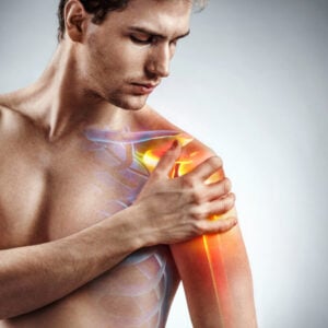
Shoulder impingement is one of the most common reasons Australians present to physiotherapy clinics. It occurs when soft tissues, primarily the supraspinatus tendon and the subacromial bursa, are compressed within the narrow space beneath the acromion as the arm is raised. This produces a dull, aching pain in the front or outer shoulder that worsens with overhead reaching, lifting, or lying on the affected side.
The evidence strongly favours conservative, non-surgical treatment for the vast majority of people with shoulder impingement. Structured physiotherapy, particularly progressive strengthening of the rotator cuff and scapular stabilisers, consistently produces outcomes equivalent to or better than surgical intervention over the long term. Most patients can return to full function without surgery.
The Anatomy Behind the Pain
The subacromial space sits between the top of the humerus and the underside of the acromion, a bony projection of the shoulder blade. The supraspinatus tendon and the subacromial bursa, a fluid-filled sac that reduces friction, pass through this space. When it narrows, these structures are repeatedly compressed during arm elevation, leading to irritation and pain.
External or subacromial impingement is the most common form, involving compression between the humeral head and the acromion. Internal impingement is more prevalent in overhead athletes. The cause may be structural, such as a hooked acromion or bone spurs, or dynamic, arising from rotator cuff weakness, poor scapular control, or glenohumeral instability. Most everyday presentations involve dynamic factors, which is why exercise-based rehabilitation is so effective.
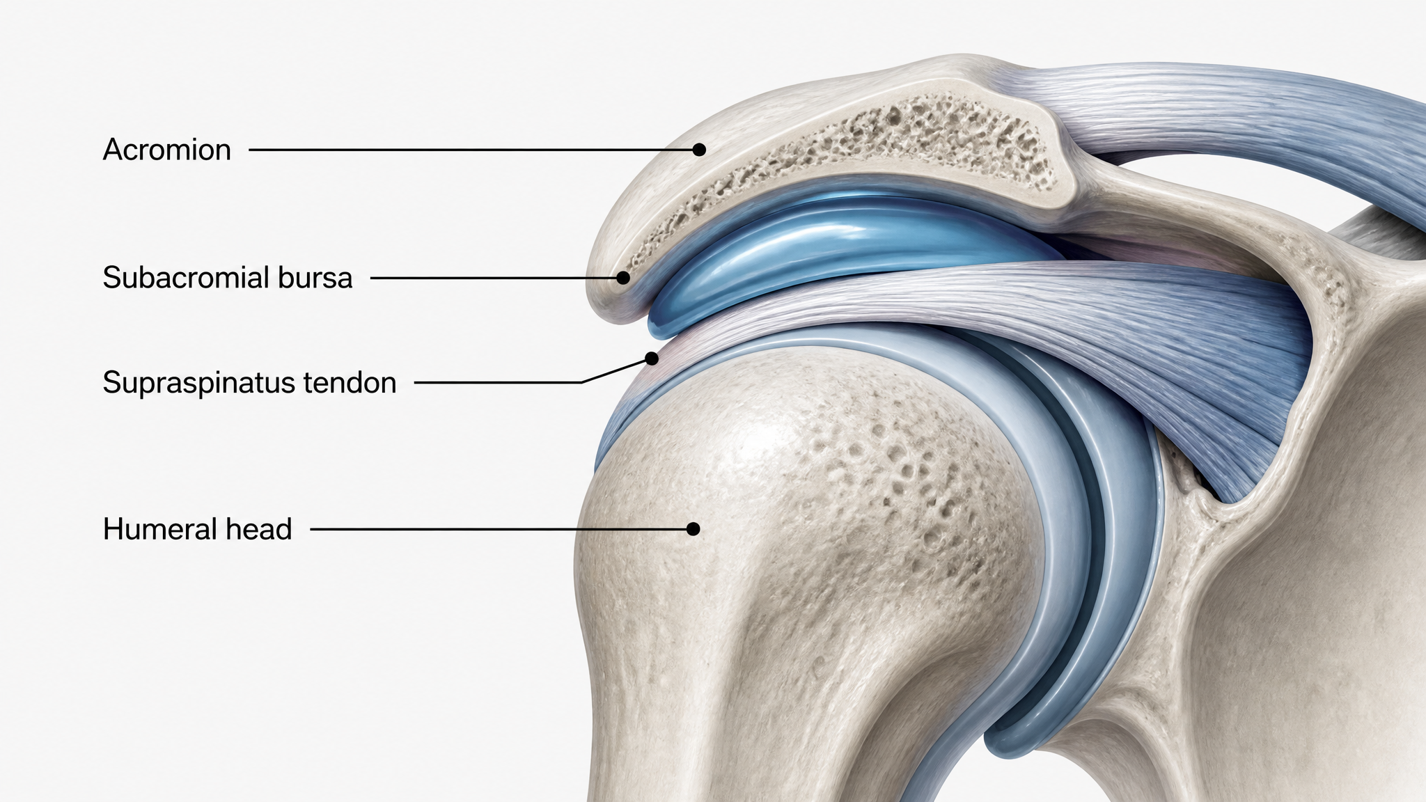
The subacromial space where impingement occurs, between the acromion above and the humeral head below
Who Is at Risk
Shoulder impingement can affect people of any age, though it becomes more prevalent as degenerative changes accumulate in the rotator cuff tendons over time. Repetitive overhead activities including painting, swimming, throwing sports, and manual trades place sustained demand on the subacromial space. Poor posture, particularly a rounded upper back, reduces the subacromial space by altering scapular position. Sudden increases in training load are another common trigger.
Diagnosis and What Clinicians Look For
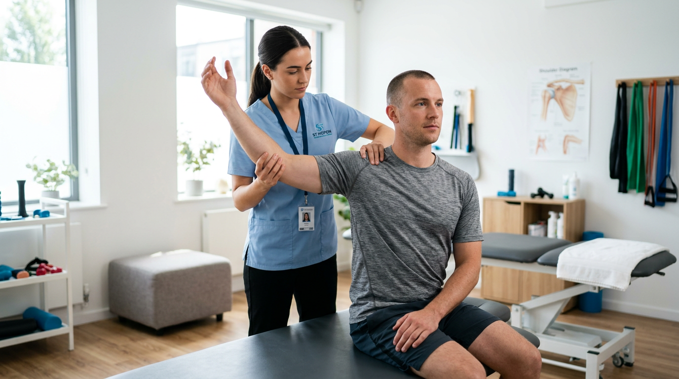
Diagnosis is primarily clinical, based on patient history and physical examination. The Neer sign involves passively flexing the arm forward in internal rotation, and pain reproduced in this position suggests subacromial involvement. The Hawkins-Kennedy test passively internally rotates the humerus with the shoulder at 90 degrees of flexion. The painful arc test identifies pain between approximately 70 and 120 degrees of shoulder abduction, the arc where subacromial compression is greatest.
Imaging is used selectively. Plain X-rays assess bony anatomy, while ultrasound or MRI is reserved for cases where symptoms persist beyond six weeks or where a significant rotator cuff tear is suspected. Structural findings on imaging do not always correlate with symptom severity, reinforcing the importance of treating the patient rather than the scan.
A Staged Approach to Rehabilitation
Modern clinical guidelines recommend categorising patients by tissue irritability before prescribing exercise intensity. The Staged Approach for Rehabilitation Classification (STAR-Shoulder) framework places patients into high, moderate, or low irritability categories, guiding how aggressively rehabilitation can progress. A patient with high irritability, significant resting pain and marked night pain, requires a gentler initial approach focused on pain reduction and rotator cuff protection. As irritability settles, the programme advances toward progressive loading.
In the early phase, submaximal isometric exercises in the scapular plane are preferred because they build rotator cuff activation without generating excessive subacromial contact pressure. Gentle range-of-motion work and manual therapy targeting the posterior capsule help restore normal glenohumeral mechanics and improve scapular positioning.
Rotator Cuff and Scapular Strengthening
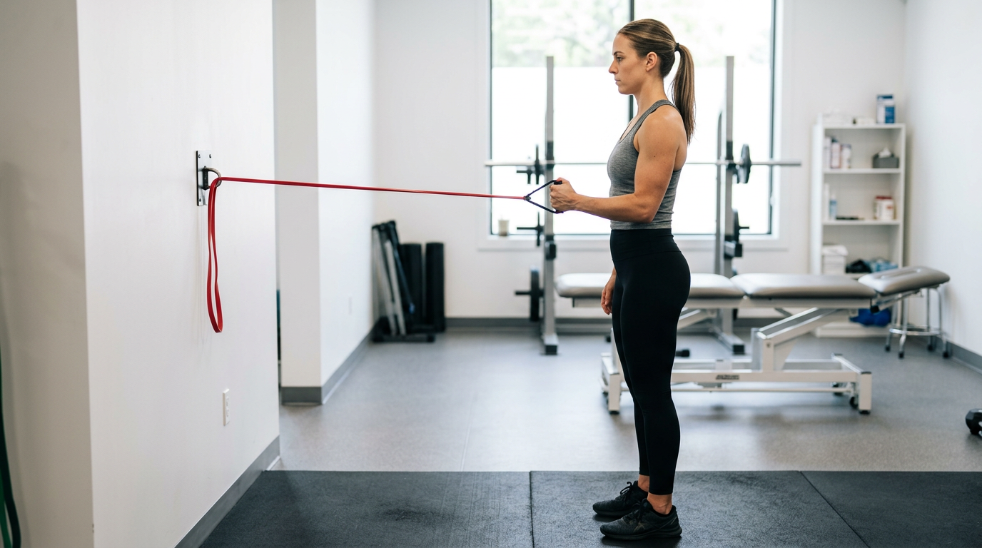
As irritability decreases, the programme shifts toward progressive strengthening. The rotator cuff functions as a dynamic stabiliser of the glenohumeral joint, keeping the humeral head centred during movement. Weakness in any of these muscles allows the humeral head to migrate superiorly, reducing the subacromial space and perpetuating impingement.
Equally important is the scapular force couple, the coordinated action of the serratus anterior and lower trapezius that rotates the scapula upward as the arm is raised. Scapular dyskinesis, characterised by poor control of the shoulder blade during arm elevation, is extremely common in people with shoulder impingement. Systematic reviews and randomised controlled trials consistently demonstrate that scapular stabilisation exercises significantly reduce pain and disability scores compared to conventional physiotherapy alone. A well-designed programme includes eccentric rotator cuff loading, serratus anterior activation, lower trapezius strengthening, and posterior capsule stretching.
Rehabilitation Phase Overview
| Phase | Primary Goals | Key Exercises | Supportive Tools |
|---|---|---|---|
| High Irritability | Pain reduction and tissue protection | Submaximal isometrics and gentle ROM | Cold therapy and kinesiology tape |
| Moderate Irritability | Restore range of motion and begin loading | Scapular setting and light band exercises | Resistance bands and heat therapy |
| Low Irritability | Strength, endurance, and return to activity | Progressive rotator cuff loading and kinetic chain work | Resistance bands and functional training aids |
What to Expect from Recovery
Most people with shoulder impingement respond well to a structured physiotherapy programme over eight to twelve weeks, though timelines vary with symptom severity and adherence to home exercises. Patient expectations and pain self-efficacy are recognised as significant prognostic indicators, often more predictive of outcomes than structural findings. Corticosteroid injections may assist when acute pain limits participation in rehabilitation but are most effective when combined with exercise.
Surgery, specifically arthroscopic subacromial decompression, is reserved for cases that have not responded to at least three to six months of well-supervised conservative management. Evidence indicates it does not produce superior long-term outcomes compared to structured physiotherapy for most patients.
A Clear Decision Summary
Shoulder impingement is a manageable condition when approached with a structured, evidence-based rehabilitation programme. Reduce irritability first, then progressively load the rotator cuff and scapular stabilisers, address the kinetic chain for active individuals, and monitor progress using functional milestones. Passive treatments and surgery rarely outperform well-supervised exercise over the long term. Consistent adherence to a quality physiotherapy programme is the most important factor in a successful recovery.
For the resistance bands, kinesiology tape, heat and cold therapy packs, and other rehabilitation tools that support shoulder recovery, browse the full range at the APE Medical Recovery and Rehabilitation collection.
References and Further Reading
This article draws on current evidence-based clinical guidance. Key sources consulted include Physiopedia (Shoulder Impingement Syndrome), the STAR-Shoulder classification framework as described in peer-reviewed physiotherapy literature, and clinical guidelines from Sports Medicine Australia. For further reading, visit Physiopedia Shoulder Impingement and Sports Medicine Australia.