
Features
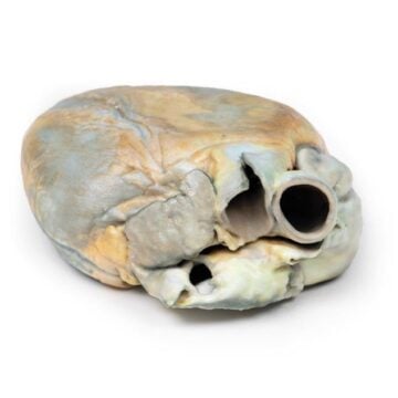
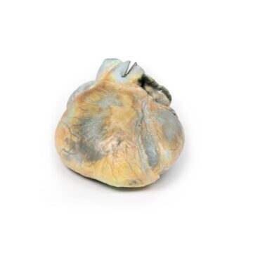
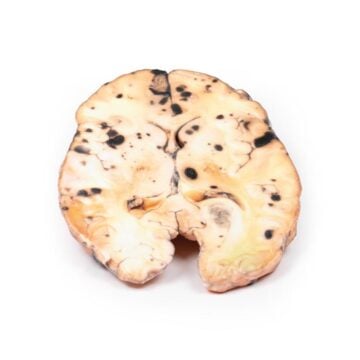
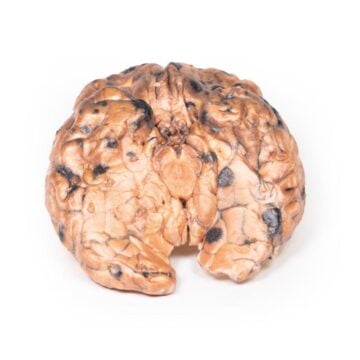
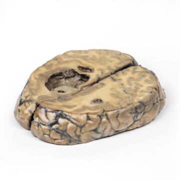
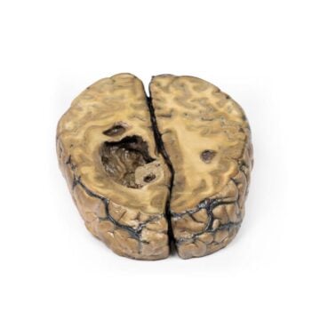
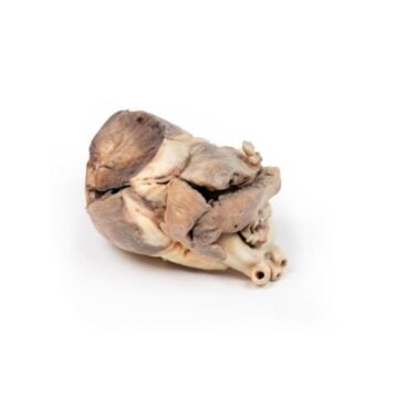
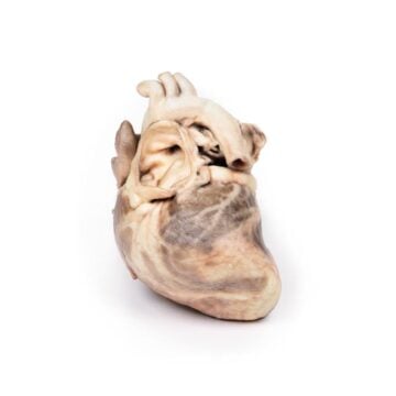
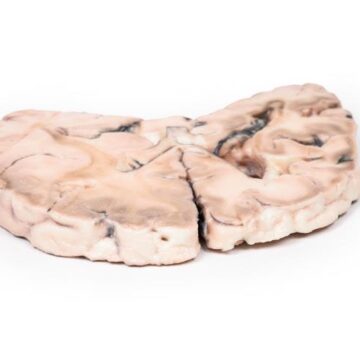
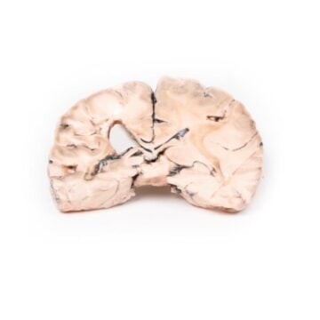
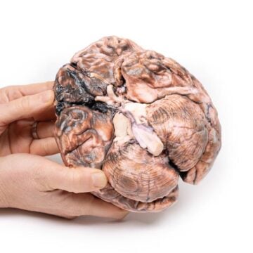
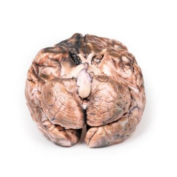
The Tertiary Syphilis is a detailed 3D pathology model created for medical educators, students, and healthcare professionals. It demonstrates the devastating advanced manifestations of syphilis with high anatomical accuracy. This model provides critical visual and tactile reference for teaching, diagnosis, and improving clinical understanding.
Examine Advanced Syphilitic Pathology Up Close with Tertiary Syphilis
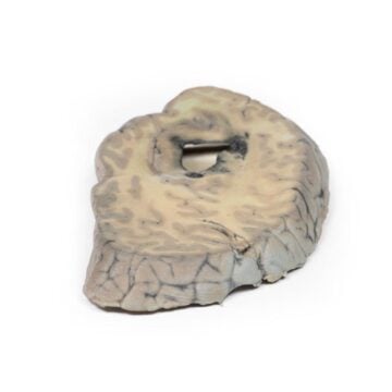
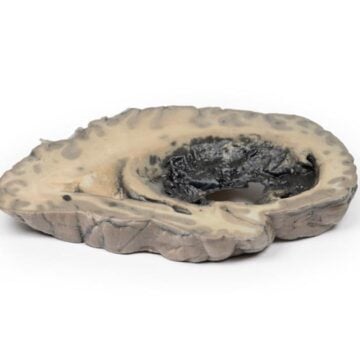
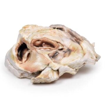
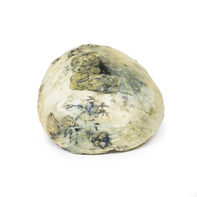
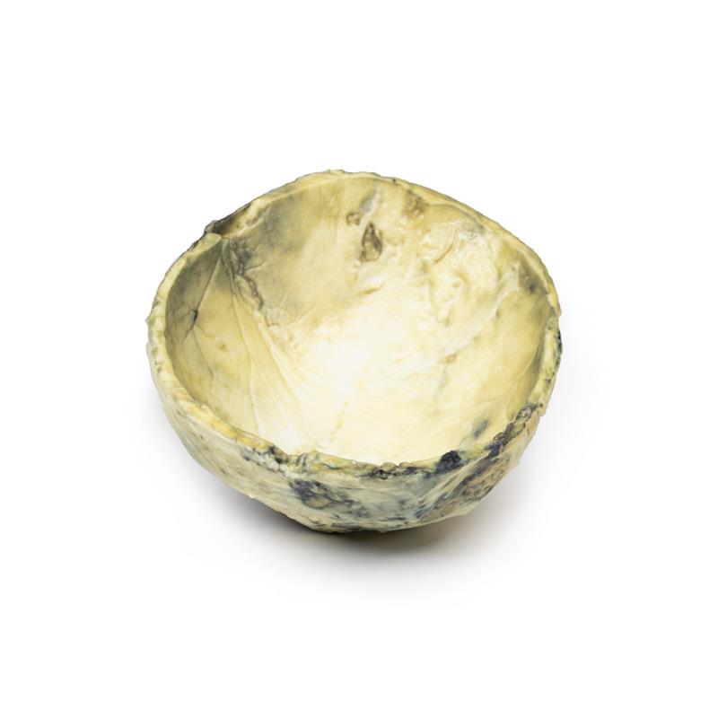
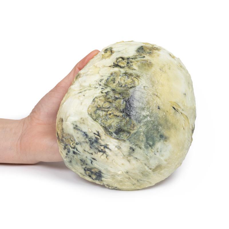
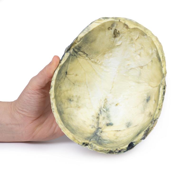
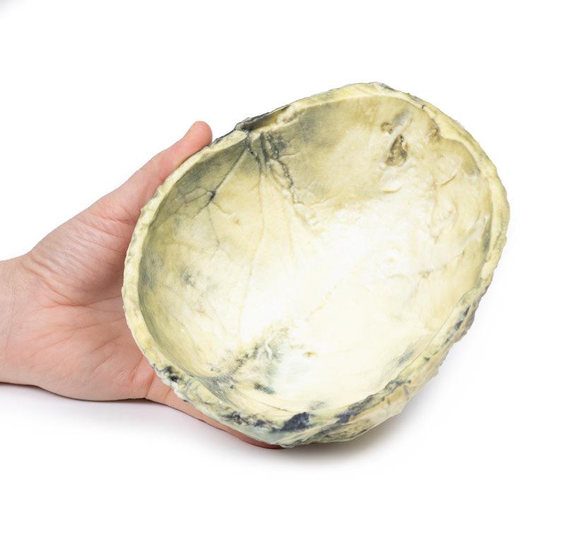
This clinical-grade Tertiary Syphilis model showcases the profound tissue destruction and characteristic lesions found in late-stage syphilitic infection. The external skull surface reveals multiple circumscribed necrotic lesions, representing gummata, with brown discoloration and sizes up to 3-4 cm. The model captures periosteal thickening and fibrinous inflammation, essential for illustrating chronic syphilitic damage. Its professional-grade build ensures long-term use and accurate disease demonstration, setting a new standard for anatomical learning about syphilis complications.
Features and Benefits
- 3D printed replica of a human skull with gummatous lesions
- Shows circumscribed necrotic areas up to 3-4 cm diameter
- Accurately highlights periosteal thickening and inflammatory changes
- Displays brown chronic syphilitic bone lesions
- Clinical history and pathological findings included for context
- Durable educational tool for medical courses and clinical environments
- Captures characteristic features of benign tertiary syphilis
Indications for Use
- Medical and pathology education
- Clinical demonstration of advanced syphilis
- Training for healthcare and diagnostic staff
- Reference for tertiary disease complications
Size Guide
- Full adult human skull vault model
- Lesion diameter – up to 3-4 cm each
- Exact anatomical dimensions of the cranial specimen represented