


Features
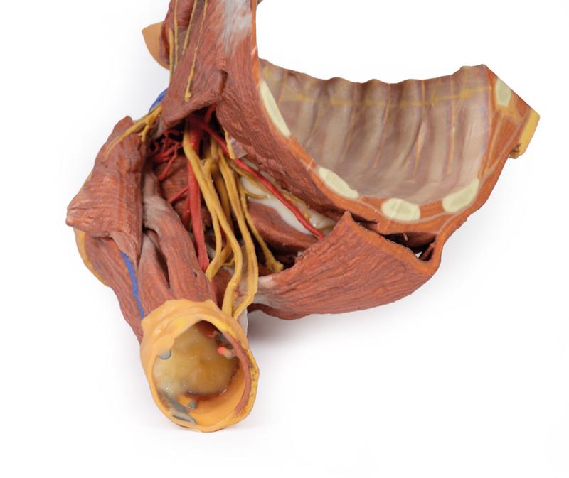
The Right thoracic wall – axilla, and the root of the neck is a premium 3D printed anatomical model designed for medical educators, students, and healthcare professionals. It provides a clear representation of the right chest wall, axilla, and root of the neck for detailed anatomical study. With this comprehensive model, users can examine essential thoracic, axillary, and cervical structures while benefiting from clinical-grade accuracy. Choose this model for precise learning and demonstration needs in physiology and medicine.
Explore Precise Anatomy with the Right thoracic wall – axilla, and the root of the neck
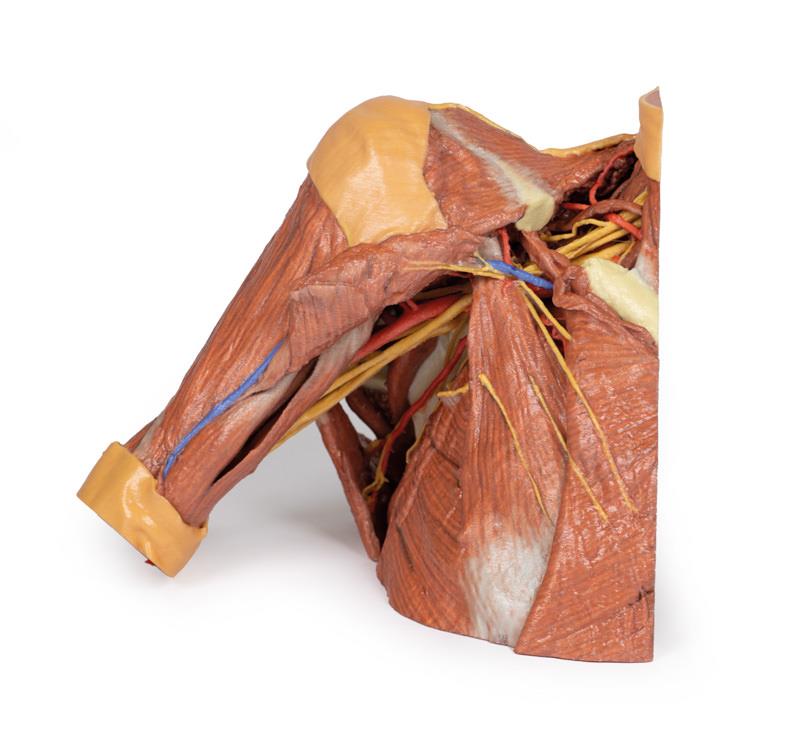
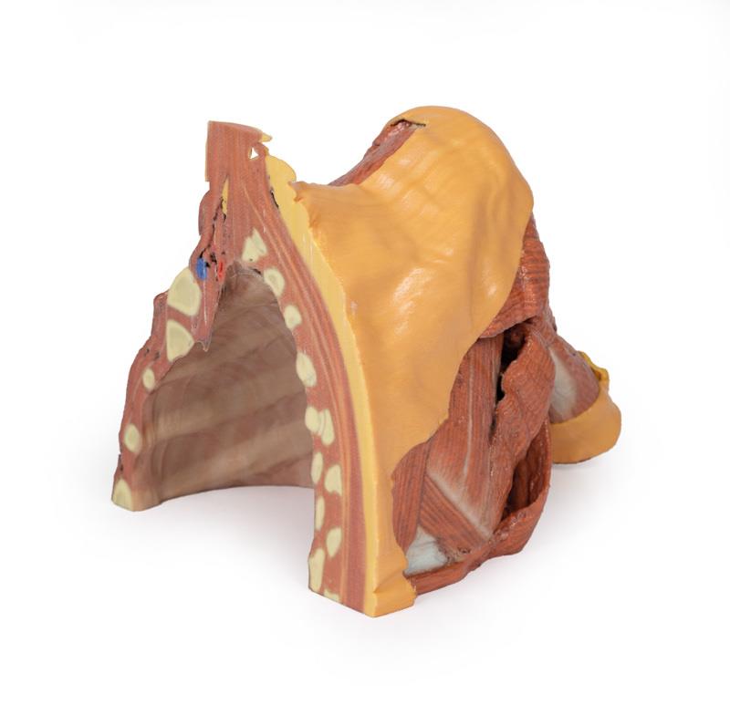
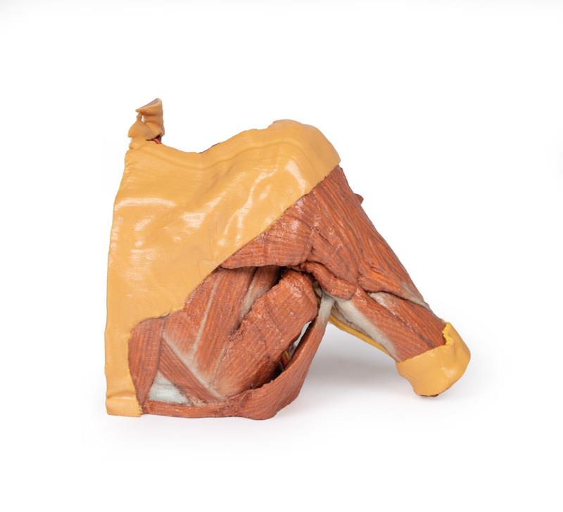
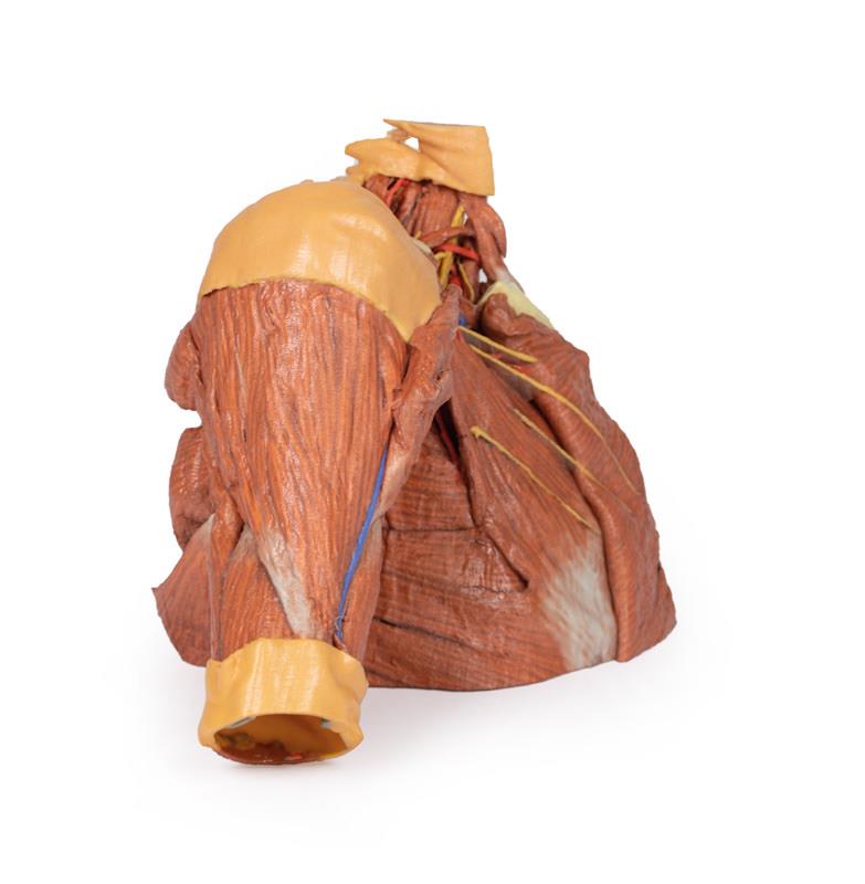
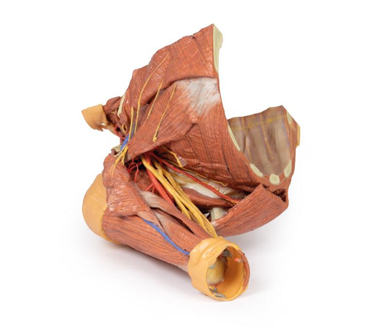
This 3D printed specimen captures the complexity of the right thoracic wall, axilla, and root of the neck, offering a valuable tool for clinical teaching and advanced anatomical coursework. The model provides direct access to bones, muscles, nerves, and vessels, making structure identification straightforward for users. An accurate parasagittal cut removes visceral contents and reveals a robust view of the parietal pleura and deep thoracic wall elements. The pectoralis major has been reflected and the pectoralis minor serves as a prominent landmark for axillary artery identification. Retained features like the subclavius muscle and near-complete brachial plexus visualization ensure a rich learning experience for professionals and students alike.
Features and Benefits
- Clinically accurate dissection with 3D printing for unparalleled realism
- Parasagittal cut enhances deep anatomical visibility
- All visceral chest contents removed for clarity
- Shows ribs and intercostal muscles beneath parietal pleura
- Full origin of neurovascular bundles visible in each intercostal space
- Pectoralis major reflected to expose pectoralis minor as a key anatomical landmark
- Middle third of clavicle removed, subclavius muscle retained
- Brachial plexus displayed nearly in its entirety from C5-T1 roots to arm entry
- Detailed anatomical description available on request
- Clinical-grade model suitable for professional education
Indications for Use
- Medical and physiotherapy teaching
- Clinical anatomy training for students and professionals
- Demonstration of thoracic, axillary, and cervical anatomy
- Hands-on anatomical study and surgical preparation
Size Guide
- One size only – full-scale 3D print of adult right thoracic wall, axilla, and root of neck specimen