

Features
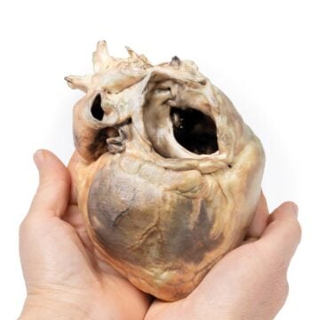
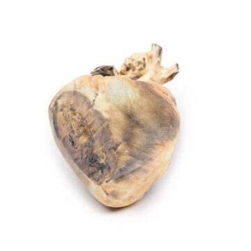
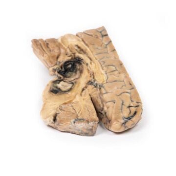
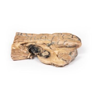
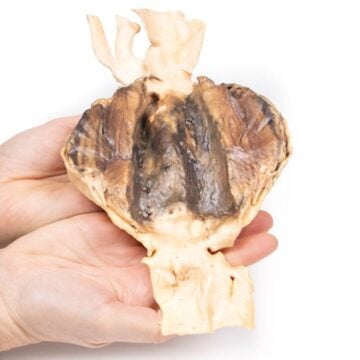
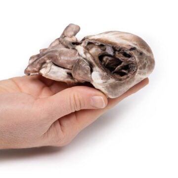
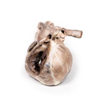
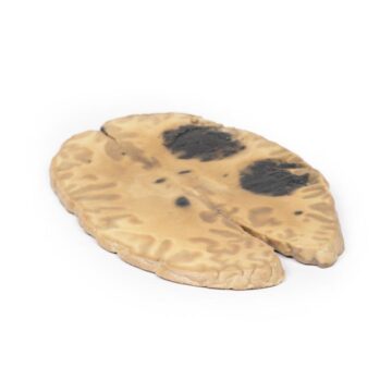
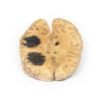
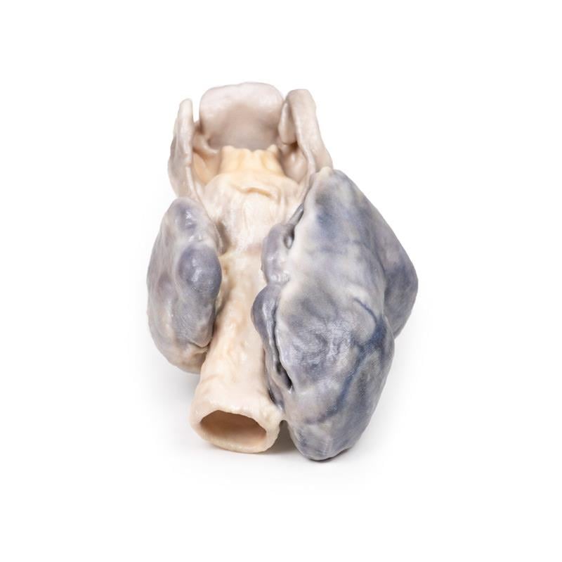
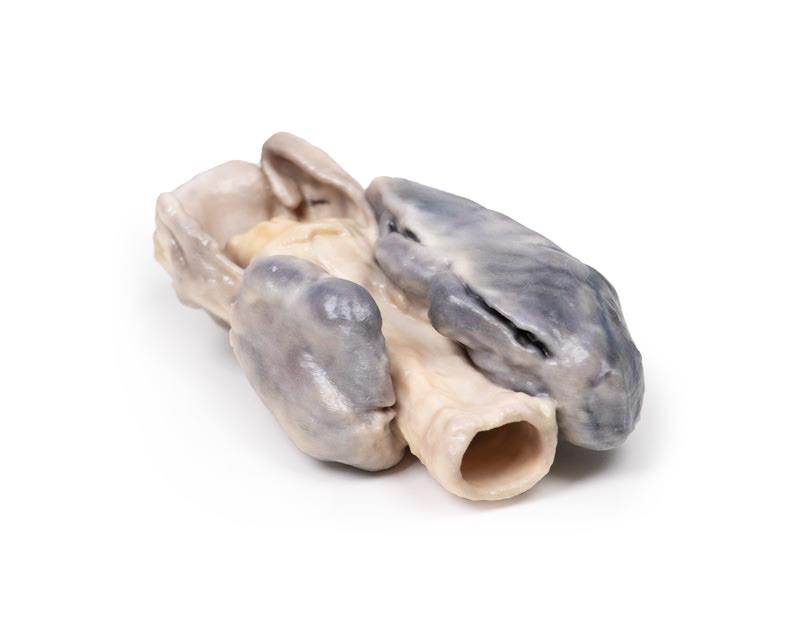
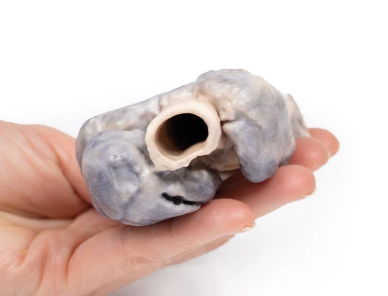
The Retrosternal Goiter is a highly detailed 3D printed anatomical model that illustrates a clinical specimen of a grossly enlarged, multilobular thyroid gland. This model helps educators and medical professionals visualize pressure symptoms and anatomical changes caused by a goiter. Choose this product for accurate, clinical-grade representation of complex thyroid anatomy, perfect for both teaching and demonstration.
Visualize Complex Thyroid Pathology with the Retrosternal Goiter Model
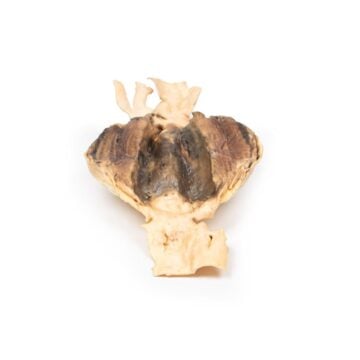
This 3D printed Retrosternal Goiter model brings critical thyroid pathology to life for students and practitioners. The model replicates the entire larynx, trachea, esophagus, and a notably enlarged thyroid, based on an actual post-mortem specimen. It reveals prominent details such as the two major lobes of the right thyroid, extended beyond usual margins, and the opened esophagus exposing the tracheal wall. The intricate anatomical features showcase abnormal growths and vascularity, enabling clear demonstration of potential pressure effects and airway impingement. The Retrosternal Goiter model provides an essential visual teaching aid for understanding goiter-related clinical presentations and complications.
Features and Benefits
- 3D printed from a real post-mortem specimen for clinical accuracy
- Displays larynx, trachea, esophagus, and enlarged multilobular thyroid
- Illustrates obvious right lobe enlargement with two distinct lobes
- Exposes posterior wall of trachea via opened esophagus
- Highlights abnormal growth and prominent surface veins
- Clinical-grade quality for teaching and demonstration
- Durable, detailed, and ready for repeated classroom or institutional use
Indications for Use
- Medical education in endocrine pathology
- Training in identifying thyroid enlargement and related symptoms
- Demonstrating effects of pressure on trachea and esophagus
- Pathology teaching for medical, nursing, and allied health students
Size Guide
- Exact anatomical scale based on original specimen
- Right thyroid lobe extends 7-8 mm beyond normal margins
- Model includes entire larynx, trachea, esophagus, and thyroid gland