


Features
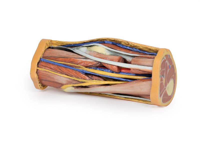
The Popliteal Fossa is a 3D printed anatomical model designed for medical students, educators, and professionals who need precise visualisation of the knee’s posterior anatomy. It reveals intricate details of the neurovascular structures and muscles, helping users study without needing cadaver specimens. Choose this model to enhance anatomical education with exceptional realism and convenience.
Unlock Clear, Accurate Anatomy with the Popliteal Fossa Model
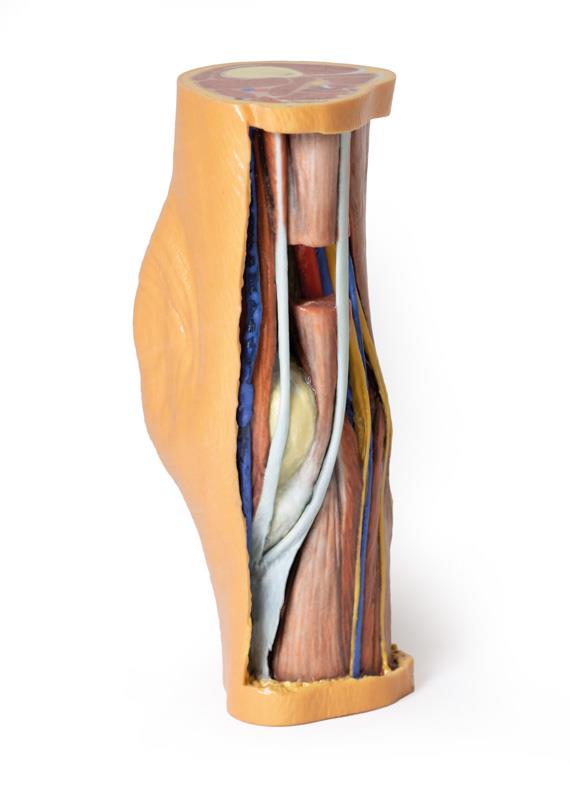
This Popliteal Fossa 3D printed model displays the distal thigh and proximal leg, expertly dissected to showcase all vital structures of the popliteal region. Remove the guesswork from anatomy lessons with lifelike reproductions of major nerves, arteries, veins, and muscle compartments. The model features both proximal and distal cross-sections, unveiling the femoral artery and vein in the adductor canal and the sciatic and great saphenous veins. A viewing window in the semimembranosus muscle provides direct access to deep vessels near the adductor magnus. All muscle borders remain intact, and the model highlights the pes anserinus, the iliotibial tract, and both superficial and deep neurovascular bundles. Enjoy ethical handling, cost savings, and consistent quality for standardized education.
Features and Benefits
- Presents distal thigh and proximal leg with posterior dissection
- Complete muscular compartments—anterior, posterior, and medial
- Femoral artery and vein visible in adductor canal
- Displays sciatic nerve and great saphenous vein
- Skin, superficial, and deep fascia removed over the fossa for maximal visibility
- Muscular borders intact with window in semimembranosus
- Exposes popliteal artery and vein near adductor magnus
- Sartorius muscle joins with semitendinosus and semimembranosus at pes anserinus
- Shows superior lateral genicular artery
- Posterior aspect of iliotibial tract descending to tibia
- Distal cross-section with ongoing nerves and vessels
- Great and small saphenous veins visible in superficial fascia
- Medial and lateral sural cutaneous nerves displayed
- Neurovascular bundles detailed in all compartments
- Accurate dimensions – 5x4x10 inches, 12-inch patellar circumference
- Printed to order with customisation options
- No ethical, storage, or regulatory complications
- Affordable and reproducible for wide educational use
Indications for Use
- Medical and physiotherapy education
- Anatomy teaching and demonstration
- Clinical reference for professionals
- Surgical training and planning
- Patient education and communication
Size Guide
- Model dimensions—5 inches (width) x 4 inches (depth) x 10 inches (height)
- Circumference around patella—12 inches