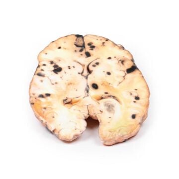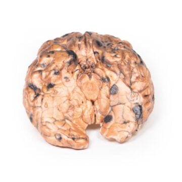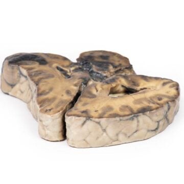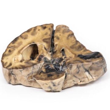
Features
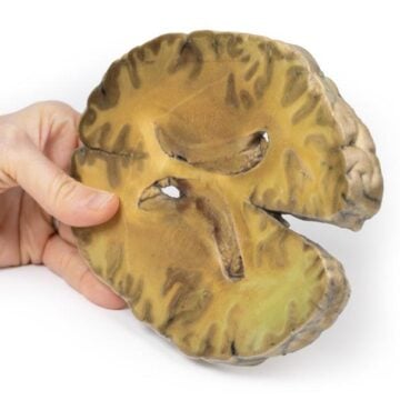
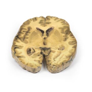
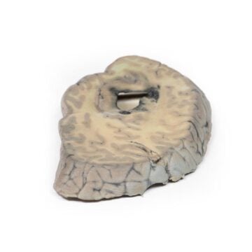
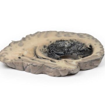
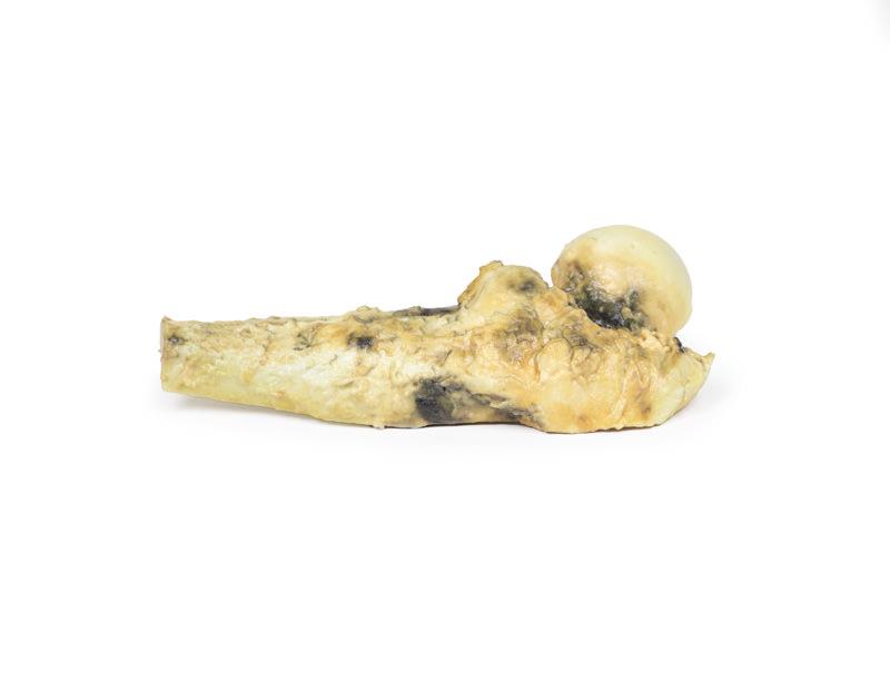
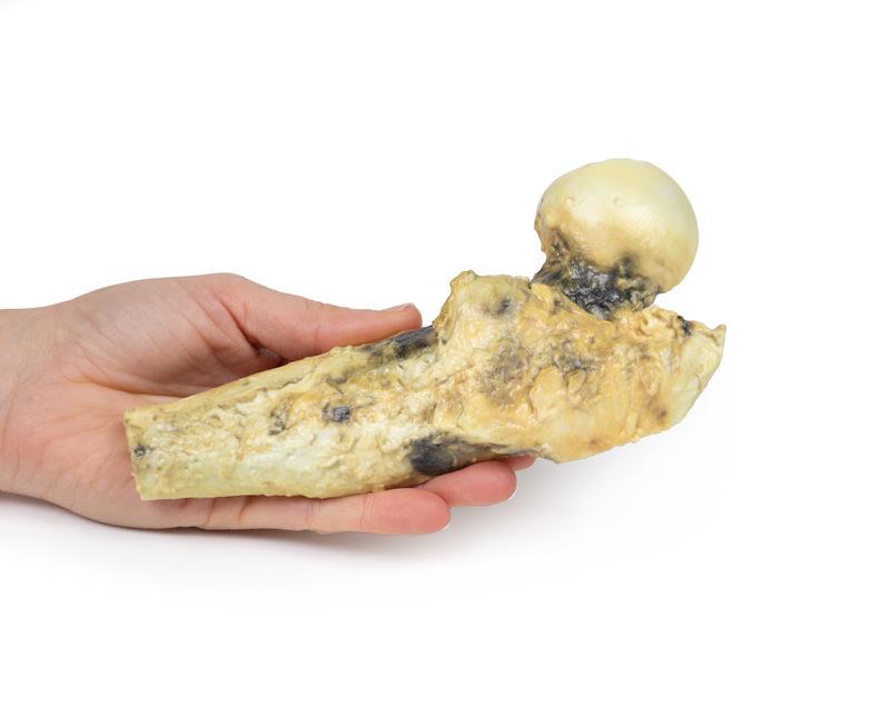
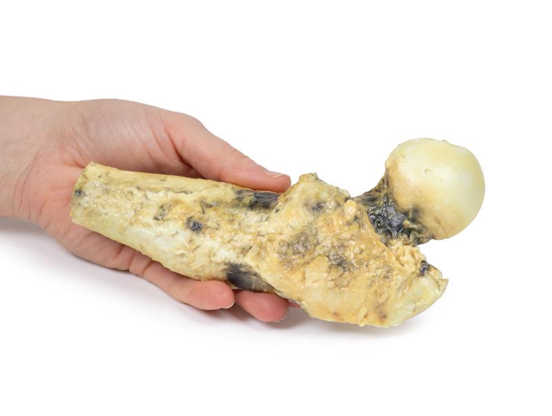
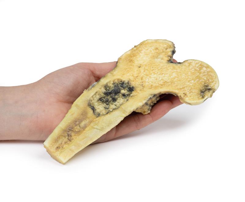
The Osteosarcoma of femur is a high-fidelity 3D printed anatomical model crafted for medical educators, students, and healthcare professionals. It provides a clinically accurate representation of a femoral tumour, supporting education in bone pathology and surgical training. This model allows users to explore the complexities of chondrosarcoma and osteosarcoma with lifelike detail not achievable through images or textbooks.
See True Bone Tumour Pathology with the Osteosarcoma of femur 3D Model
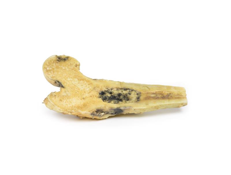
This clinical-grade model replicates the head, neck, and upper third of the right femur, including a realistic 6.5 cm ovoid tumour within the medullary cavity. You can view the specimen longitudinally sawn to reveal a haemorrhagic, pale hyaline, and cystic tumour. The Osteosarcoma of femur model demonstrates the changes seen on X-ray and CT, such as bony absorption, periosteal reaction, and tumour expansion. Educators and trainees gain a hands-on opportunity to study the appearance and context of malignant femoral tumours, improving anatomical understanding and surgical planning. Choose this model for its unmatched precision and value in explaining tumour progression and resection challenges.
Features and Benefits
- 3D printed for clinical-grade anatomical accuracy
- Displays head, neck, and upper third of right femur
- Presents an ovoid, 6.5 cm tumour in the medullary shaft
- Shows realistic pathological features – haemorrhagic, hyaline, and cystic areas
- Sawn longitudinally to allow internal inspection
- Ideal for teaching, demonstration, and surgical simulation
- Supports CT and X-ray correlation for case-based learning
Indications for Use
- Medical and anatomy education focusing on bone tumours
- Surgical planning and simulation for femoral tumour excision
- Demonstration of osteosarcoma and chondrosarcoma pathology
- Patient and trainee education regarding malignant bone lesions
Size Guide
- Replicates the head, neck, and upper third of the right femur
- Tumour maximum diameter – 6.5 cm (ovoid lesion in shaft)
- Full specimen dimensions match adult male femur anatomy