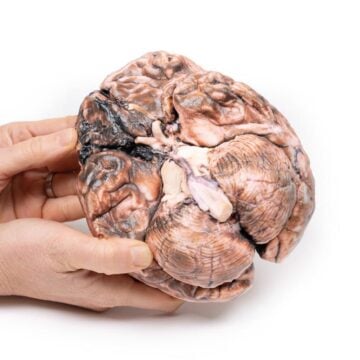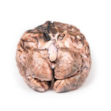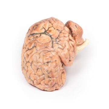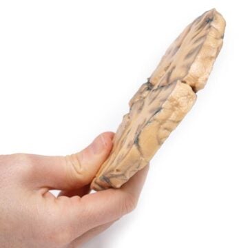

Features
The Osteochondroma is a clinically-accurate 3D printed anatomical model designed for medical professionals, educators, and students. It provides a realistic representation of a common benign bone tumor, supporting clear understanding and improved diagnosis. Choose the Osteochondroma model for precise pathology demonstration and superior educational value in orthopedics and oncology.
Enhance Medical Education with a Detailed Osteochondroma Model
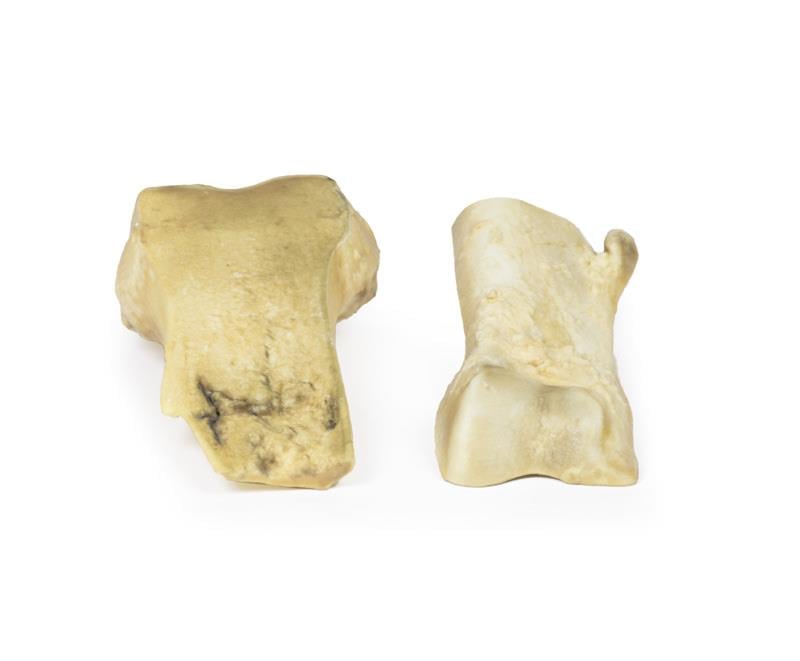
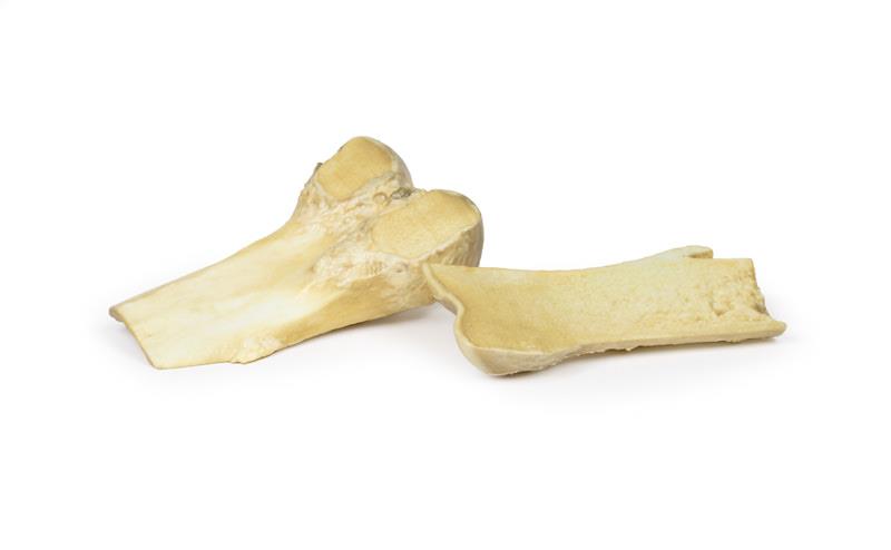
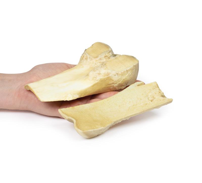
This Osteochondroma 3D printed model replicates the lower end of a human femur, featuring a 2 cm pedunculated bony outgrowth capped with hyaline cartilage. The model illustrates the typical presentation of osteochondroma, including its location 7 cm above the medial condyle of the femur. Instructors and practitioners gain a tactile resource that aids in teaching the clinical appearance, structural characteristics, and pathology of this benign tumor. Built to high clinical standards, this model ensures robust use for classroom and surgical training. It supports recognition of osteochondroma and aids in understanding complications such as neurovascular compression and fracture risk. Durable construction and exceptional detail make this model a vital educational tool.
Features and Benefits
- Clinically-accurate 3D printed model of an osteochondroma on the femur
- Shows a 2 cm pedunculated lesion with hyaline cartilage cap
- Displays external surfaces as seen in real pathology specimens
- Educational resource ideal for orthopedics, pathology, and oncology training
- Durable and high-quality construction for repeated handling
- Helps visualize bony protuberance and anatomical relationships
- Supports teaching on bone tumor diagnosis and treatment options
Indications for Use
- Medical education on benign bone tumors
- Demonstration of osteochondroma pathology during clinical and academic lectures
- Hands-on training for medical students and health professionals
- Orthopedic and oncology case studies and discussions
- Aid in patient education about osteochondroma
Size Guide
- Model represents a femoral specimen with a bony projection 2 cm in length
- Projection located 7 cm above the medial condyle on the medial aspect of the femoral shaft
- Exact anatomical proportions based on clinical case details