Features
The Metastatic Tumour in Lung from Primary Testicular Cancer is a precision 3D printed anatomical model developed for advanced education in pathology and oncology. It offers medical professionals and students a realistic view of lung metastasis from testicular cancer, providing a critical resource for understanding tumour spread and complex disease presentations. Choose this model to deliver accurate, hands-on teaching for real-world clinical scenarios and improve pathology learning outcomes.
Transform Pathology Education with a Clinical-Grade 3D Lung Metastasis Model
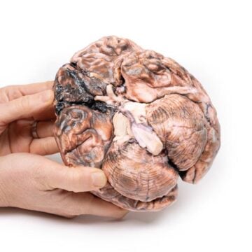
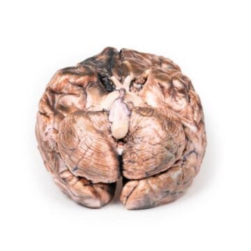
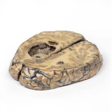
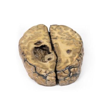
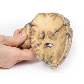
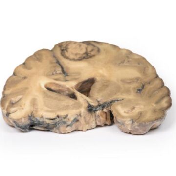
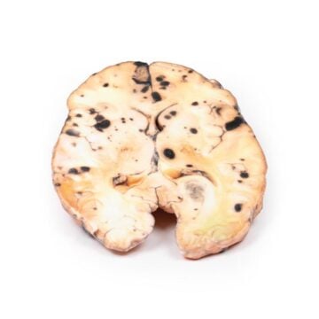
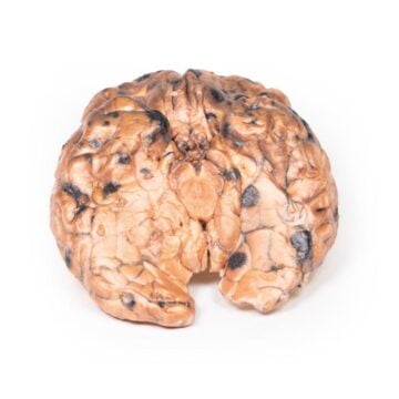
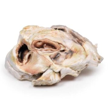
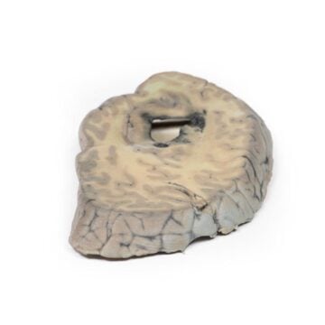
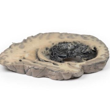
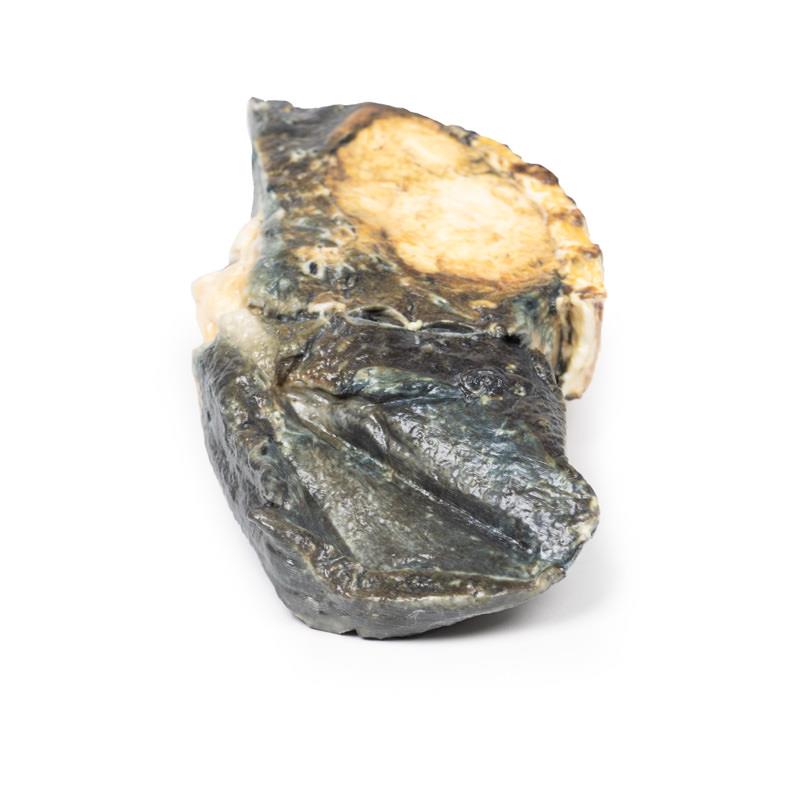
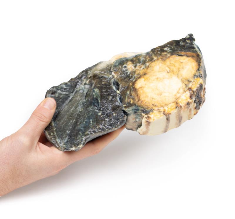
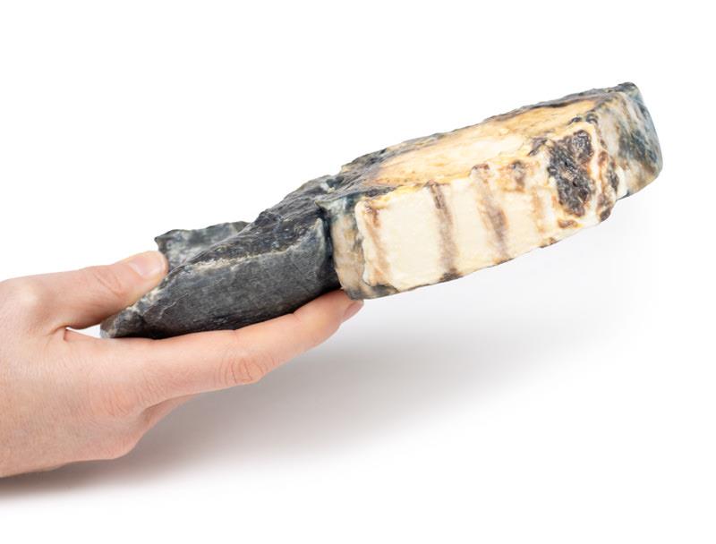
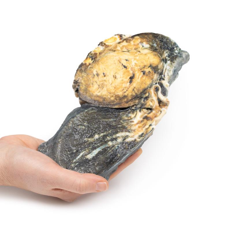
This exceptionally detailed Metastatic Tumour in Lung from Primary Testicular Cancer model presents an authentic replica of a right lung invaded by mixed germ cell tumour metastases, including portions of four ribs. The model displays multiple rounded tumour nodules in lung parenchyma, ranging from 5 to 30 mm, with variegated coloration and regions of necrosis and haemorrhage. One tumour casts the lower lobe bronchus, while others protrude from the pleural surface. You experience an unrivaled, tactile representation for teaching, clinical demonstration or pathology exam preparation, offering clear insights into pulmonary metastasis from testicular origins.
Features and Benefits
- Highly detailed 3D printed model based on a true clinical case
- Depicts a right lung with metastatic mixed germ cell tumours
- Displays multiple nodules, some up to 30 mm in diameter
- Shows realistic features such as necrosis, haemorrhage, and tumour casts
- Includes portions of four ribs for anatomical context
- Designed for educational, research, and clinical training use
- Clinical-grade quality for accurate teaching
- Manufactured by The Anatomy Store, trusted by Australian medical institutions
- Durable and suitable for repeated handling
Indications for Use
- Medical and pathology education focusing on metastatic testicular cancer
- Student examination and hands-on teaching in medical schools
- Clinical demonstration of complex tumour spread
- Professional training for oncologists, pathologists, and radiologists
Size Guide
- Full right lung model including four attached rib segments
- Multiple tumour nodules from 5 mm to 30 mm in diameter featured
- Life-size replication based on clinical specimen