Features
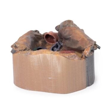
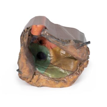
The Heart and the distal trachea, carina and primary bronchi is a high-fidelity 3D printed anatomical model crafted for medical educators, students, and clinicians. It provides an authentic representation of the heart and crucial airway structures, making complex anatomical relationships clear. This model supports advanced teaching, demonstration, and surgical training by offering anatomy that is difficult to visualise in textbooks or generic models.
Explore Realistic Cardiac and Airway Anatomy in the Heart and the distal trachea, carina and primary bronchi Model
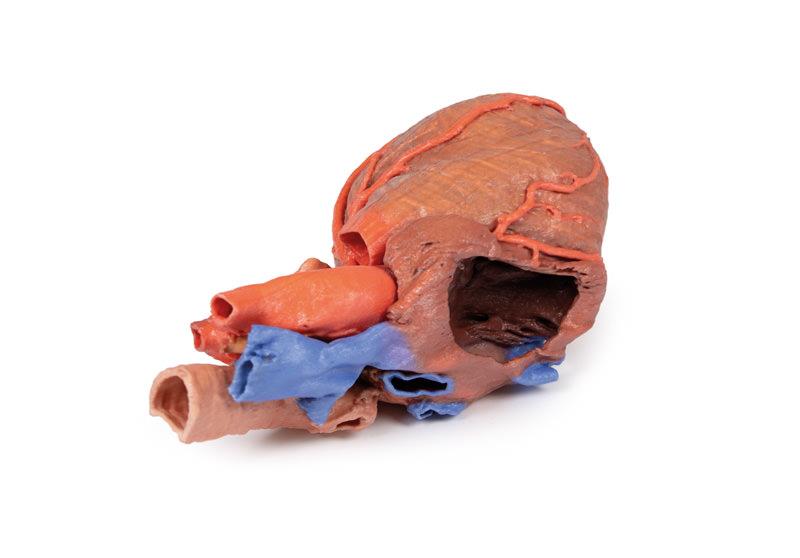
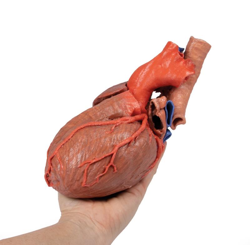
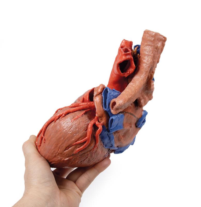
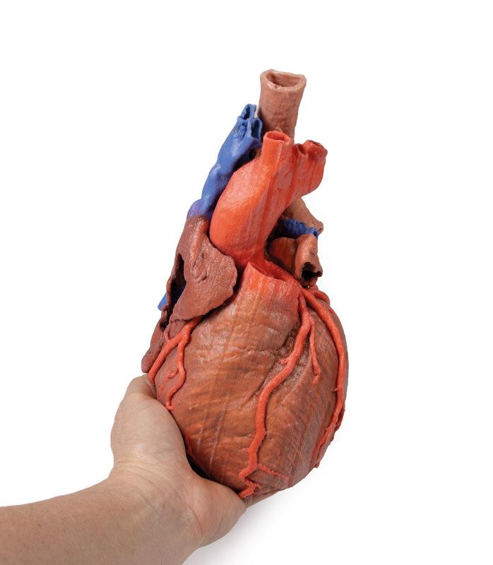
This expertly printed specimen brings together the external anatomy of the heart, the distal trachea, carina, and primary bronchi within the posterior mediastinum. Vividly preserved great vessels and the left atrium, along with the pericardial reflections of both transverse and oblique sinuses, ensure thorough anatomical accuracy. An anterior window reveals the tricuspid valve and access to the right ventricle, while both coronary arteries and their branches remain visible for an in-depth study. With the left auricle sectioned and the circumflex artery course revealed, users can examine coronary circulation up close. The open pulmonary valves and intact aortic arch display the origins of vital branches. The model omits the cardiac veins but retains the coronary sinus. Preservation of the terminations of the brachiocephalic and azygos veins into the superior vena cava completes the educational experience.
Features and Benefits
- Precisely 3D printed from real anatomical data for lifelike detail
- Displays the external heart and airway structures within the posterior mediastinum
- Shows great vessels, left atrium, pericardial reflections, and sinuses
- Anterior window exposes right atrium interior, base of auricle, and tricuspid valve
- Both right and left coronary arteries and branches are clearly visualised
- Sectioned left auricle demonstrates circumflex artery course in coronary groove
- Pulmonary trunk is removed, offering view of open pulmonary semilunar valves
- Arch of aorta remains intact to show principal arterial branches
- Coronary sinus preserved, aiding venous anatomy studies
- Terminations of left/right brachiocephalic veins and azygos vein into SVC maintained
Indications for Use
- Medical teaching in cardiology, pulmonology, and anatomy courses
- Surgical and procedural demonstration or training
- Patient education in clinical settings
- Reference for complex anatomical relationships during case discussions
Size Guide
- One standard specimen size – accurately reflects adult cardiac and mediastinal proportions