Features
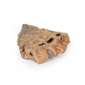
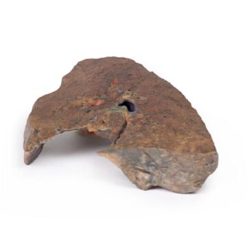
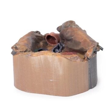
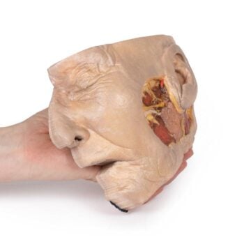
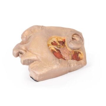
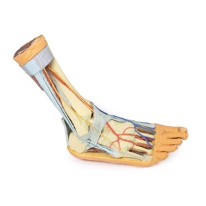
The Foot – Superficial and deep structures of the distal leg and foot is a precise 3D printed anatomical model crafted for healthcare educators, clinicians, and students. It enables in-depth demonstration of both superficial and deep anatomy of the right distal leg and foot. Choose this model to visualize anatomical relationships that textbooks cannot show, enhancing both teaching and clinical understanding.
Reveal Deep and Superficial Anatomy Clearly with This Advanced 3D Model
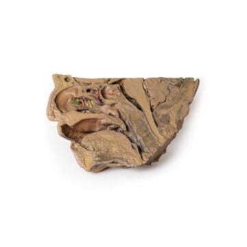
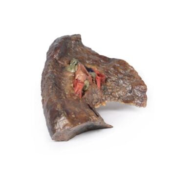
Unlock unmatched detail during your anatomy sessions with the Foot – Superficial and deep structures of the distal leg and foot. This 3D printed model reveals superficial and deep compartments, demonstrating muscles, nerves, arteries, veins, ligaments, and tendons in their true spatial relationships. Easily follow the tibial nerve and posterior tibial artery, see the preserved origin of the great saphenous vein, and observe detailed ligament structures on the lateral aspect. Made to order, this model serves educators and clinicians who need accurate, three-dimensional insights for effective hands-on anatomy explanation and clinical examination instruction.
Features and Benefits
- Shows both superficial and deep structures of the right distal leg and foot
- Posterior compartment features deep muscles with triceps surae and tendocalcaneous removed
- Clear paths of tibial nerve and posterior tibial artery, branching to medial and lateral plantar regions
- Origin of abductor hallucis brevis removed for better nerve and artery visualization
- Preserved origin of great saphenous vein from the medial dorsal venous arch ascending through the specimen
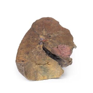
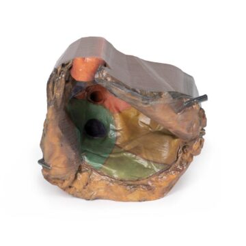
- Anterior compartment muscles removed to display interosseous membrane, anterior tibial artery, and deep fibular nerve
- Tendinous insertions of tibialis anterior, extensor hallucis longus, and extensor digitorum longus retained
- Continuity of anterior tibial artery through dorsalis pedis to arcuate and dorsal metatarsal arteries shown
- Dorsal interosseous muscles removed to reveal terminal branches to plantar interosseous muscles
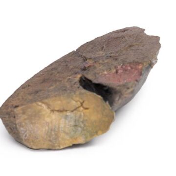
- Lateral aspect displays fibularis longus and brevis muscles and their tendons
- Details insertion of fibularis brevis, termination of superficial fibular nerve, and abductor digiti minimi origin
- Exposes key ligaments including anterior/posterior tibiofibular, calcaneofibular, talonavicular (dorsal/posterior), and deltoid ligament
- Clinical-grade detail for education and examination
- 3D printed to order for custom, up-to-date representation
Indications for Use
- Anatomy education for students, clinicians, and educators
- Demonstration of anatomical relationships in medical training
- Clinical skill teaching for examination and diagnosis
- Surgical training support and anatomical review
Size Guide
- Single model representation of right distal leg and foot in life-size dimensions
- Exact anatomical proportions preserved based on medical imaging data
- No alternative sizes available; custom print ensures unique, true-to-life scaling