")

")
Features
Advanced endoscopic transphenoidal surgery simulator.
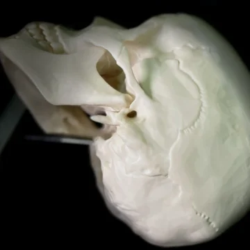
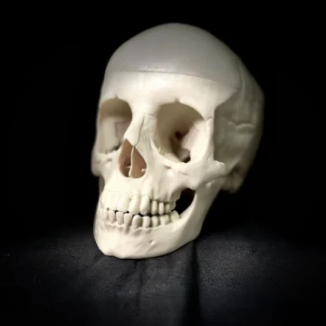
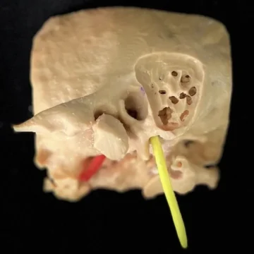
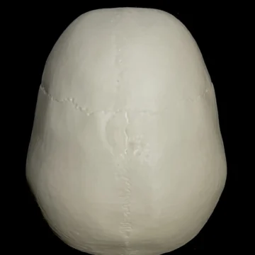
Human skull with all the craniometric points, fissures and canals. 1:1 scaled from a real human CT scan with similar drilling properties to the human bone. Includes skin, nasal mucosa, turbinates, septum, as well as a detailed intrasphenoidal anatomy.
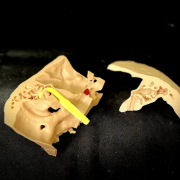
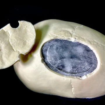
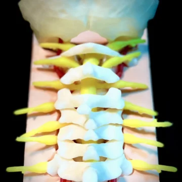
The inner skull is covered with dura, and includes all cranial nerves, intracranial arteries as well as the brain, cerebellum and brainstem. Pathology included: pituitary adenoma and planum sphenoidale meningioma extracted from real cases and including the DICOM images for the preoperative planning.
The ‘Endonasal Endoscopic Simulator’ is designed to perform any endonasal approach under a precise skull base anatomy.
All the different components of this simulator are made from real DICOM images extracted from CT scans and Magnetic Resonance images.
The skin, skull, meninges, neural structures, vessels as well as the tumors, are manufactured to provide them with similar properties to the real ones and recreate the most realistic sensations.
The simulator also includes the preoperative DICOM files to allow the user studying each individual case, carry out a detailed surgical planning and perform the surgery itself.
Expand your endonasal surgery in the sagittal and coronal planes to gain space, and enjoy the anatomical journey!