Features
The Craniocervical Junction Surgery Simulator is a clinical-grade model designed for surgeons, educators, and trainees who require realistic practice with advanced anterior and posterior approaches. It replicates the full anatomical complexity of the craniocervical junction, giving you hands-on training that elevates surgical skills and confidence. This simulator enables you to plan and execute procedures in a controlled, risk-free environment.
Elevate Surgical Training with the Most Realistic Craniocervical Junction Model
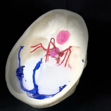
Push your surgical expertise further with the Craniocervical Junction Surgery Simulator. Created directly from a real human CT scan, this simulator delivers 1 -1 scaled anatomy featuring detailed occipital bone, cervical vertebrae, vertebral arteries, intervertebral discs, and neural elements. You experience bone-like resistance while drilling, mimicking both cortical and cancellous bone. Practise complex operations including anterior cervical discectomy and fusion, corpectomy, posterior cervical discectomy, laminectomy, and screw fixation. The model displays disc herniation pathology, making it ideal for mastering every step from planning screw entry points to executing precise surgeries. Use this tool to simulate both anterior and posterior approaches with unmatched anatomical accuracy.
Features and Benefits
- Human 3D-printed craniocervical junction with lifelike anatomical detail
- Made from real human CT scan data at a 1 -1 scale
- Mimics human cortical and cancellous bone drilling properties
- Includes occipital bone, cervical vertebrae, vertebral arteries, intervertebral discs, spinal cord, and nerves
- Integrates disc herniation pathology for realistic case scenarios
- Supports a wide range of surgical exercises and approaches
- Perfect for planning precise screw entry and trajectory
- Delivers risk-free, repeatable practice for surgeons and trainees
Indications for Use
- Simulation of anterior and posterior craniocervical junction approaches
- Training for anterior cervical discectomy and fusion
- Corpectomy practice
- Posterior cervical discectomy
- Laminectomy and screw fixation technique
- Education on disc herniation pathology management
Size Guide
- 1 -1 scale direct from a real human CT scan
- Includes full occipital bone and cervical vertebrae segments (no size options)
- One anatomical size representing standard adult anatomy