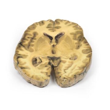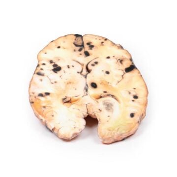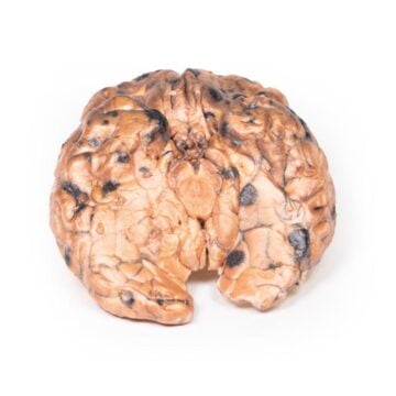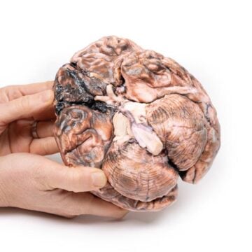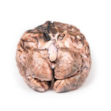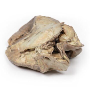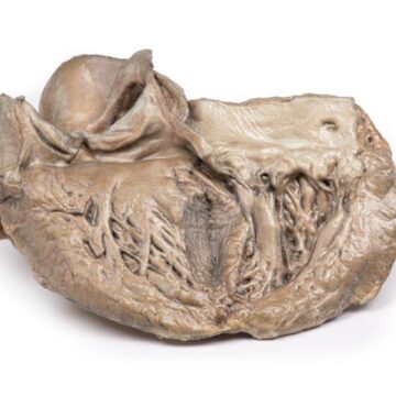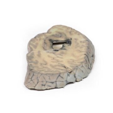

Features
The Carcinoma of Larynx is a detailed 3D printed anatomical model designed for medical educators, students, and healthcare professionals. It demonstrates the advanced pathology of laryngeal cancer, supporting both clinical teaching and in-depth study. This educational tool helps clarify complex anatomy and disease progression, offering a precise resource for training and examinations.
Enhance Medical Learning with the Carcinoma of Larynx Model
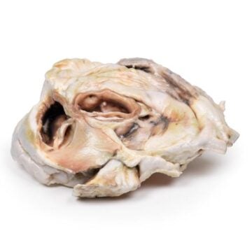
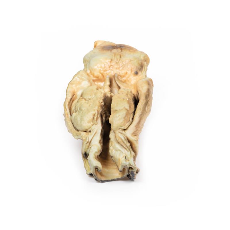
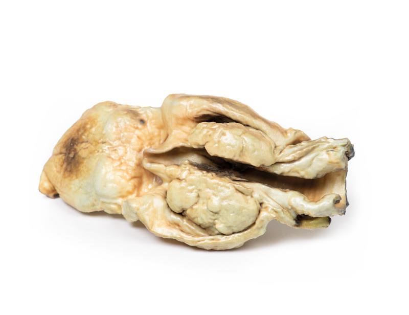
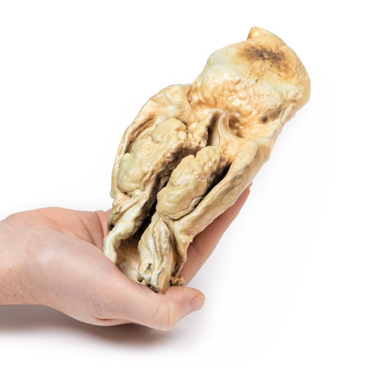
This clinical-grade Carcinoma of Larynx model brings real-pathology education into the classroom and training environment. The model features a mounted specimen depicting tongue, pharynx, larynx, oesophagus, and trachea in the coronal plane. It displays a 5 x 4 x 2 cm fungating carcinoma extending into both pyriform fossae with distinct necrotic and irregular surfaces. The cancer invades vocal cords, the left aryepiglottic fold, and both pyriform fossae, offering clear visualization for learners to understand tumour spread and anatomy. Proven helpful for recognising risk factors and common symptoms, this model also aids in illustrating squamous cell carcinoma progression and impact. Choose this 3D printed teaching aid for accuracy, durability, and impactful demonstrations in any medical classroom or training session.
Features and Benefits
- High-fidelity 3D printed anatomical specimen
- Coronally mounted with tongue, pharynx, larynx, oesophagus, and trachea
- Detailed depiction of a 5 x 4 x 2 cm fungating carcinoma
- Shows tumour involvement of both vocal cords, left aryepiglottic fold, and pyriform fossae
- Educational focus on tumour necrosis, irregularity, and anatomical relationships
- Clinical-grade resource for accurate pathology representation
- Ideal for teaching risk factors, clinical presentation, and disease progression
Indications for Use
- Medical, nursing, and allied health education
- Clinical training and examination scenarios
- Pathology and oncology instruction
- Anatomy teaching for undergraduate and postgraduate students
- Professional development for healthcare providers
Size Guide
- Specimen size 5 x 4 x 2 cm carcinoma displayed within the full laryngeal, pharyngeal, oesophageal, and tracheal anatomy; mounted in the coronal plane