
Features
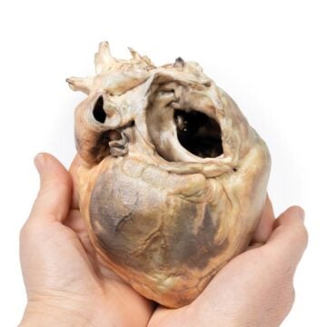
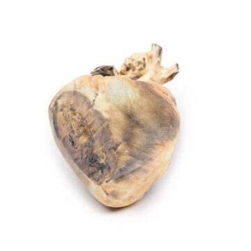
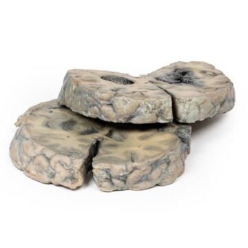
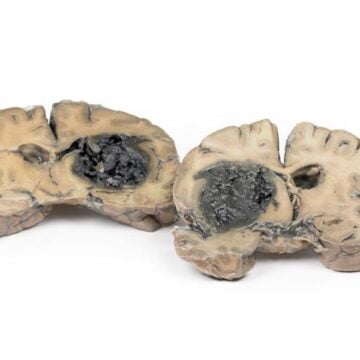
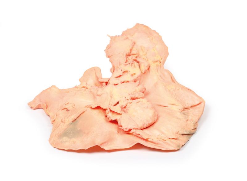
The Adenocarcinoma of the stomach is a highly detailed 3D printed anatomical model designed for medical educators, students, and healthcare professionals. It provides realistic insight into the pathology and features of gastric adenocarcinoma, supporting accurate teaching and advanced study. This model helps users visualize critical disease patterns and complications, ensuring a deeper understanding of gastric cancer for diagnostic and educational purposes.
Deepen Your Medical Knowledge with the Adenocarcinoma of the stomach 3D Model
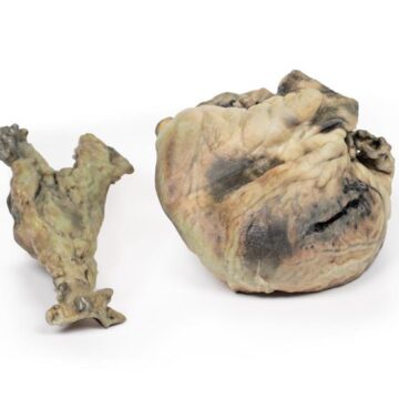
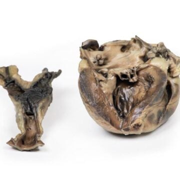
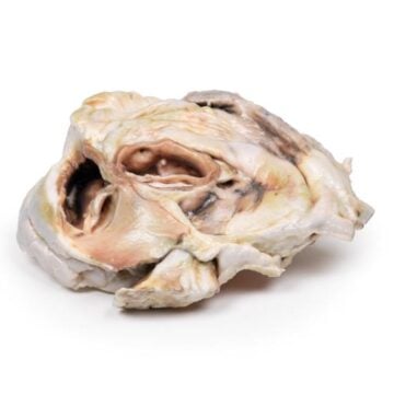
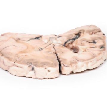
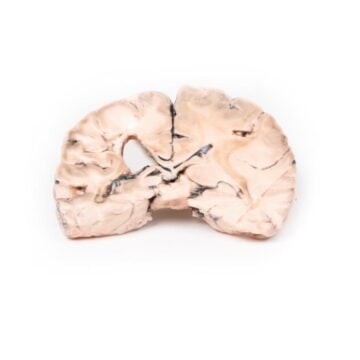
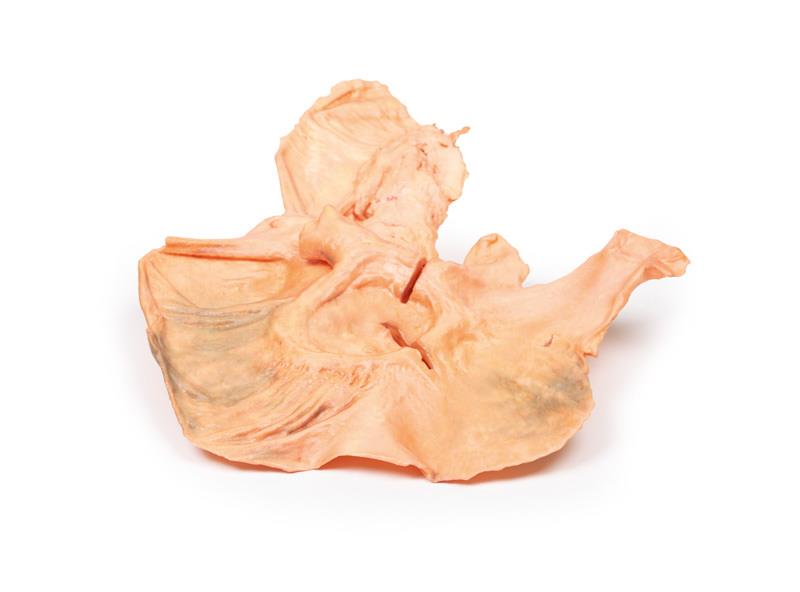
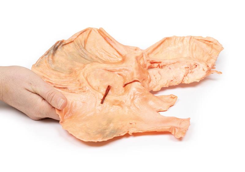
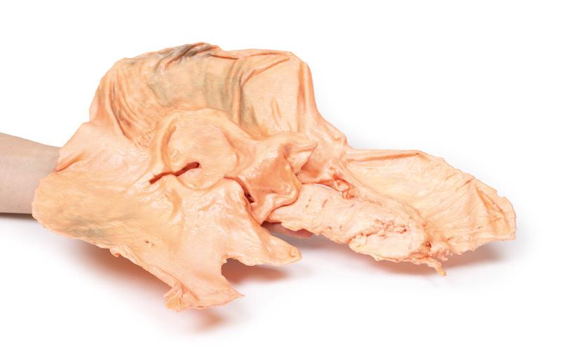
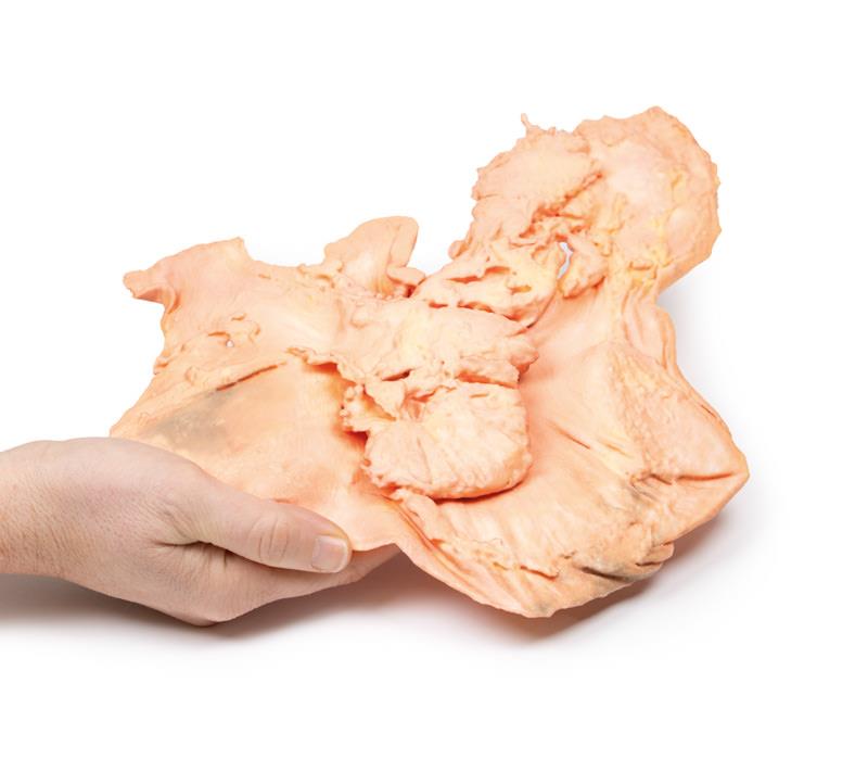
This clinical-grade model delivers a lifelike sagittal slice of the oesophagus, stomach, proximal duodenum, and pancreas. The Adenocarcinoma of the stomach model highlights key pathological elements – a 7x5cm ulcer on the lesser curve, radiating rugae loss, raised edges, and necrotic debris. The specimen reveals ulcer edge elevation by tumor tissue, two eroded arteries with recent hemorrhage, and pancreas adhesion, matching true disease progression. Histology sites on the model demonstrate well-differentiated adenocarcinoma and direct invasion, supporting robust educational exploration. Geographically accurate, with realistic representation of both intestinal and diffuse types, this resource assists in identifying risk factors and understanding disease morphology.
Features and Benefits
- Precision 3D printed, clinical-grade anatomical model
- Sagittal view includes oesophagus, stomach, duodenum, and pancreas
- Represents large 7x5cm ulcer on lesser curve
- Raised, rolled ulcer edges with necrotic base and rugae loss
- Realistic depiction of tumor elevation and recent hemorrhage
- Adhesion of pancreas to ulcer for accurate visualization
- Clearly marked histology sampling sites (3cm defects)
- Demonstrates tumour invasion into adjacent structures
- Ideal for medical teaching, pathology labs, and specialist instruction
Indications for Use
- Medical education and training
- Pathology demonstration and advanced study of gastric cancers
- Visualization of clinical and post-mortem findings
- Support for lectures, self-study, and examination preparation
- Training on gastric ulcer and adenocarcinoma identification
Size Guide
- Full sagittal section anatomical model showing entire stomach and adjacent organs
- Ulcer dimension – 7 x 5 cm
- Histology defects – 3 cm diameter each