Features
Transform Your Neuroanatomy Education with Remarkable Accuracy
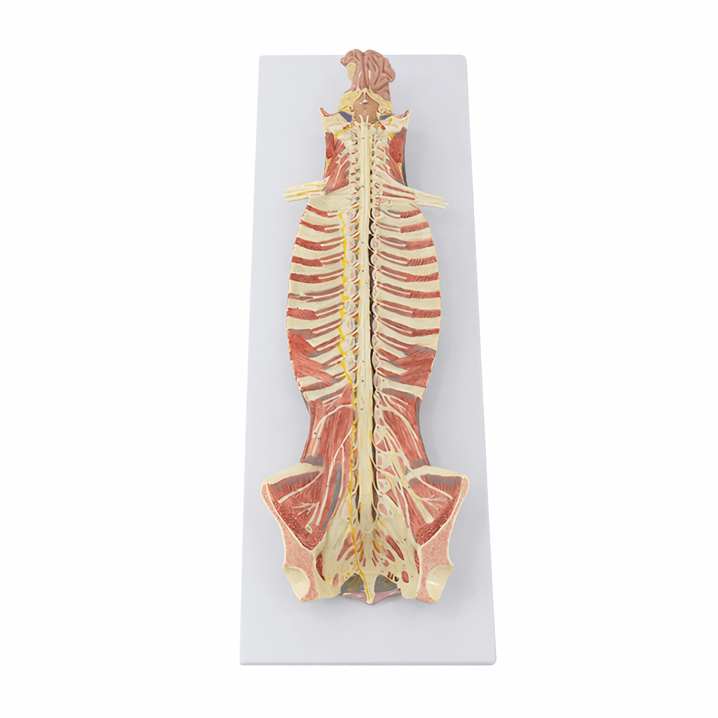
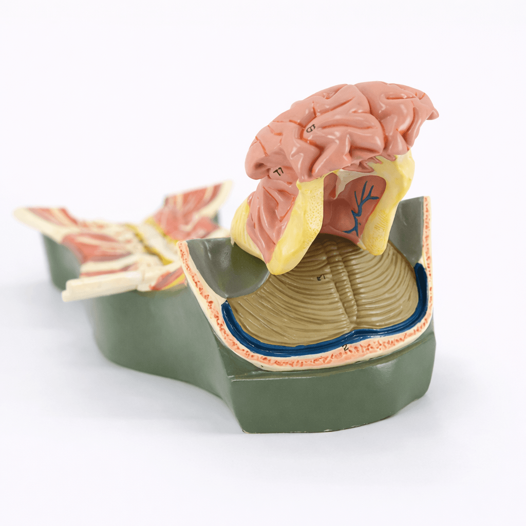
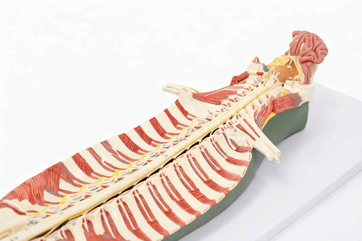
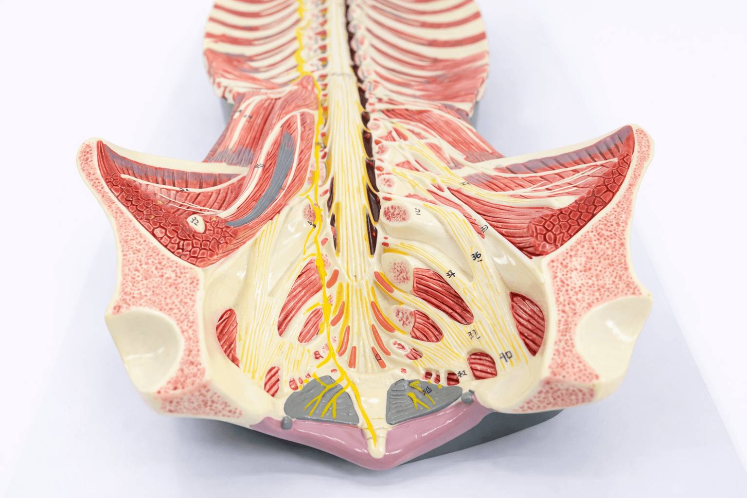
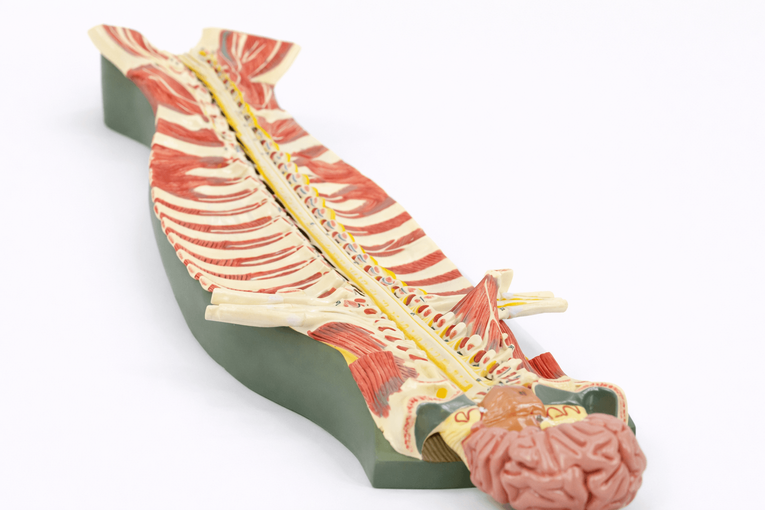
Bring your teaching and learning sessions to life with the Spinal Cord in Vertebral Canal Model. This detailed, durable neuroanatomy model enables hands-on exploration of the spinal cord and vertebral canal relationship. Examine nerve roots, ganglia, and dura in lifelike relief. Spinal nerves and vertebrae appear in accurate alignment, helping educators and students visualize anatomical structures clearly. The Spinal Cord in Vertebral Canal Model creates an immersive classroom experience, strengthening memory retention and anatomical comprehension. Crafted for longevity and realism, it serves medical students, physiotherapists, and healthcare professionals who demand accuracy and durability in anatomical demonstration. Easy to transport, this model facilitates engaging presentations in clinical settings, lectures, and patient education. Elevate your educational tools and inspire trust with precise, tactile illustration of spinal cord anatomy.
Features and Benefits
- Displays detailed spinal cord and vertebral canal relationship
- Accurately represents nerve roots, ganglia, and dura mater
- Shows clear spinal nerve emergence from each vertebra
- Durable, high-quality construction ensures longevity
- Lifelike colors improve anatomical identification
- Removable from base for hands-on study
- Lightweight and easy for transport between educational settings
- Ideal for classrooms, clinics, and patient demonstrations
Indications for Use
- Medical and physiotherapy education
- Anatomical demonstration for students and patients
- Clinical instruction for healthcare professionals
- Patient education in spinal cord injuries and disorders
Size Guide
- Model length 42 cm
- Model width 8 cm
- Model height 6 cm