
Features
The Stages of Disc Degeneration is a clinical-grade anatomical model created for health professionals, educators, and students. It demonstrates spinal disc pathology progression, making complex spinal conditions clear and accessible. Choose this model to offer hands-on learning for teaching or patient education using true-to-life physical examples.
Discover Spinal Pathologies with the Stages of Disc Degeneration Model
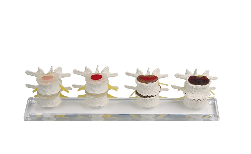
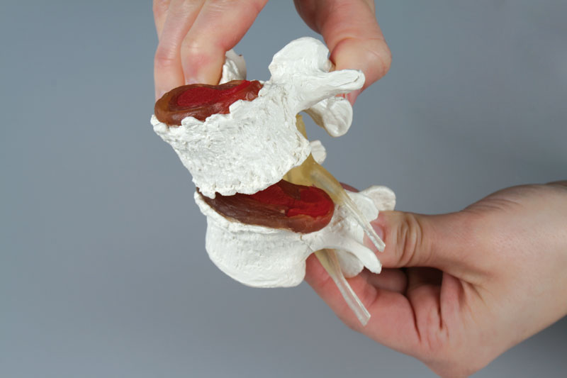
This detailed model showcases four paired vertebrae in sequence, presenting both healthy and pathological conditions. You see a healthy disc, slight disc damage, disc prolapse with bone degeneration, and advanced bone and disc degeneration, all mounted magnetically on a sturdy plexiglass base. Each pair detaches effortlessly for interactive demonstration and examination. The flexible vertebrae construction allows users to display realistic spinal movements, providing an engaging, tactile experience. Clinical-grade quality and compact dimensions make this a versatile choice for teaching clinics, classrooms, and patient consultations. Rely on this model for vivid, physical illustration of spinal health and degeneration.
Features and Benefits
- Four pairs of vertebrae show the normal state and three pathologies
- Magnetic mounting ensures easy removal from plexiglass base
- Flexible vertebrae allow movement demonstration
- High-quality, clinical-grade construction
- Perfect for education, demonstration, and patient engagement
- Clear progression from healthy to advanced degeneration
- Lightweight at 0.9 kg for easy transport and handling
Indications for Use
- Medical and anatomy education for students and professionals
- Patient education in clinics and consultations
- Spinal condition demonstration and pathology progression
- Physical therapy and rehabilitation explanation tools
Size Guide
- Model length – 38 cm
- Model width – 10 cm
- Model height – 10 cm
- Weight – 0.9 kg