Features
The Renal Cell Carcinoma is a 3D printed anatomical model designed for medical professionals, students, and educators. It showcases the detailed pathology of renal cell carcinoma, demonstrating tumor involvement, metastasis, and associated structural changes in the kidney. This model helps users understand clinical features and typical presentation, making it ideal for medical training and patient education.
Elevate Medical Education with a High-Fidelity Renal Cell Carcinoma Model
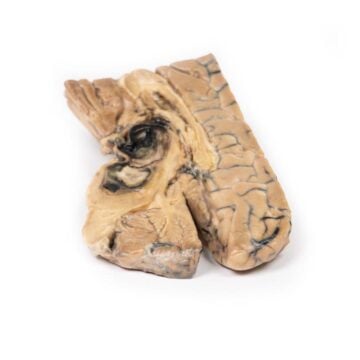
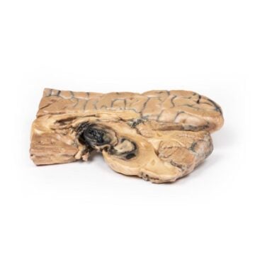
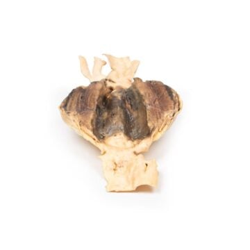
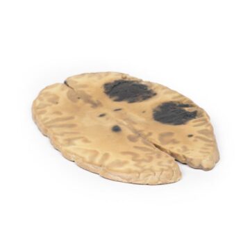
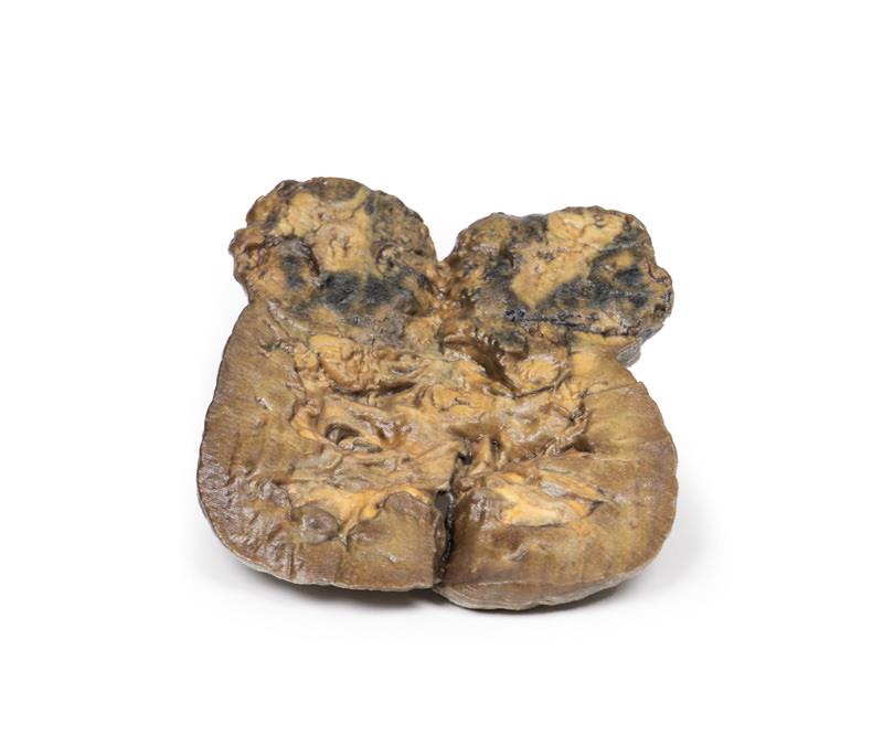
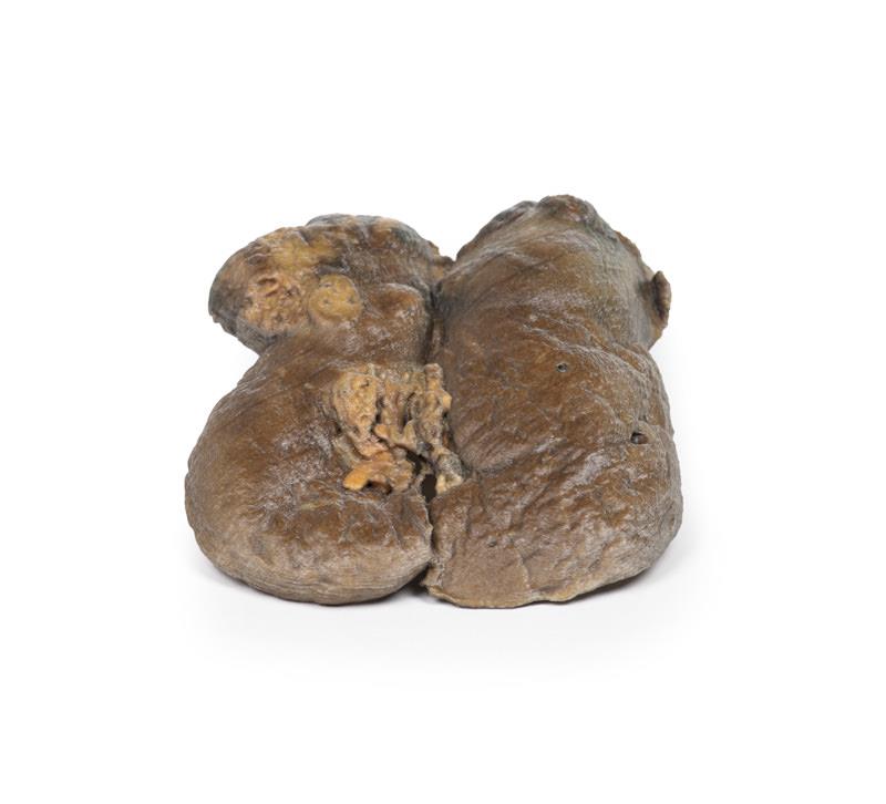
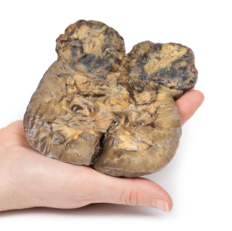
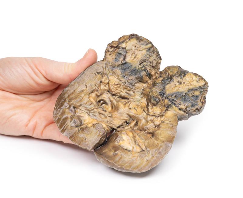
This advanced 3D printed Renal Cell Carcinoma model brings the reality of kidney cancer into the classroom and clinical settings. The model presents a kidney dissected in the coronal plane, revealing a large, irregular malignancy in the lower pole, along with multiple metastatic nodules. Varied colors and textural differences highlight regions of hemorrhage, necrosis, and cystic changes. You can examine hydronephrosis, papillary blunting, and distinct capsular features, including scars and cysts. Instructors and students use this model to visualize complex disease progression, understand anatomical relationships, and improve diagnostic recognition. Clinical-grade quality ensures an accurate representation for teaching or demonstration in any setting.
Features and Benefits
- Precision 3D printing displays real-life tumor size, shape, and variegated surface
- Includes intrarenal metastases with small pale nodule formations
- Models hemorrhage, necrosis, capsular nodularity, and cystic structures on the kidney
- Demonstrates hydronephrosis with dilated pelvis and blunted papillae
- Clearly shows tumor-induced distortion and compression of renal tissue
- Mounted for optimal viewing of cut surface anatomy
- Clinical-grade detail for accurate pathology education
Indications for Use
- Medical education for renal carcinoma pathology
- Training for clinicians, students, and patient education sessions
- Demonstration of metastatic spread within the kidney
- Anatomy teaching in universities or hospital-based programs
Size Guide
- Model represents an adult human kidney
- Lower pole tumor measures 5 cm in diameter
- Displays intrarenal metastatic nodules above and separate from the main mass
- Life-size anatomical proportions for accurate study and reference