Features
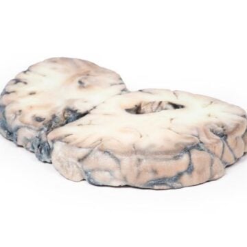
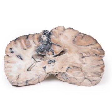
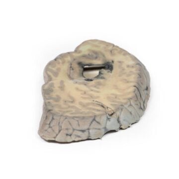
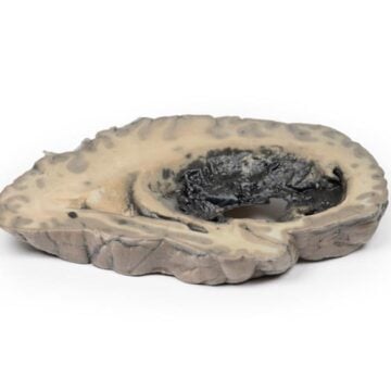
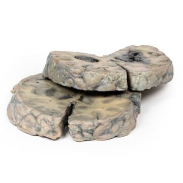
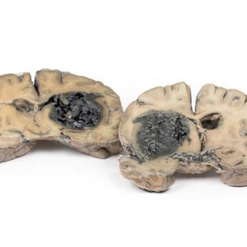
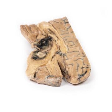
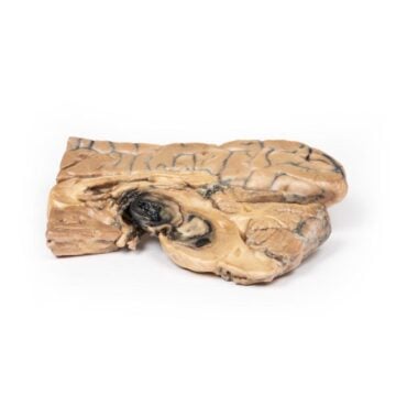
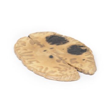
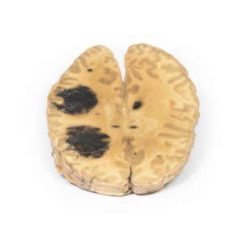
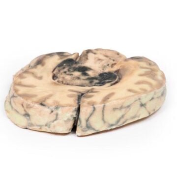
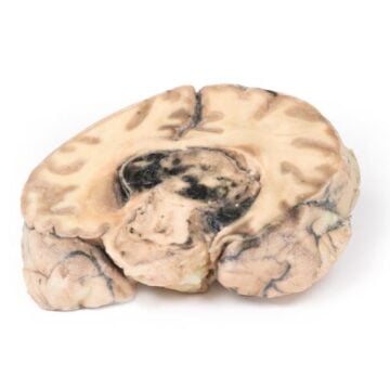
The Lung Multiple Secondary Carcinoma Deposits in The Lung and Pleura is a clinical-grade 3D printed anatomical model created for medical educators and students. It illustrates the spread and pathology of metastatic carcinoma within the lung and pleura, providing a highly visual, hands-on teaching solution. Choose this model to demonstrate complex pulmonary metastasis scenarios with exceptional detail that enhances medical learning and discussion.
Reveal the Complexities of Pulmonary Metastasis with a True-to-Life 3D Model
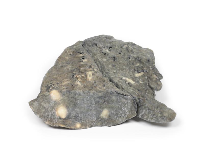
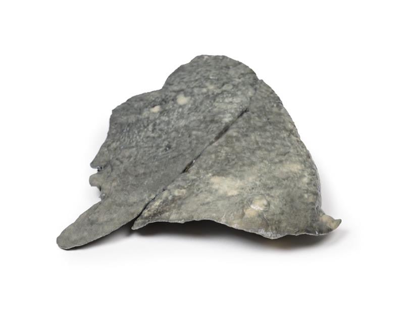
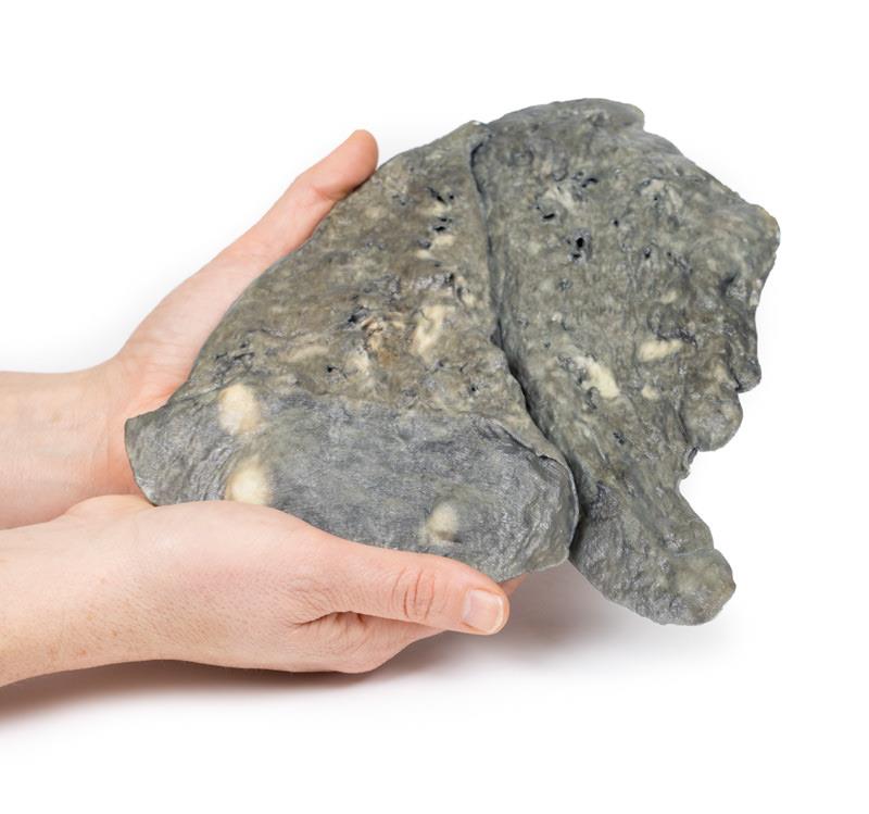
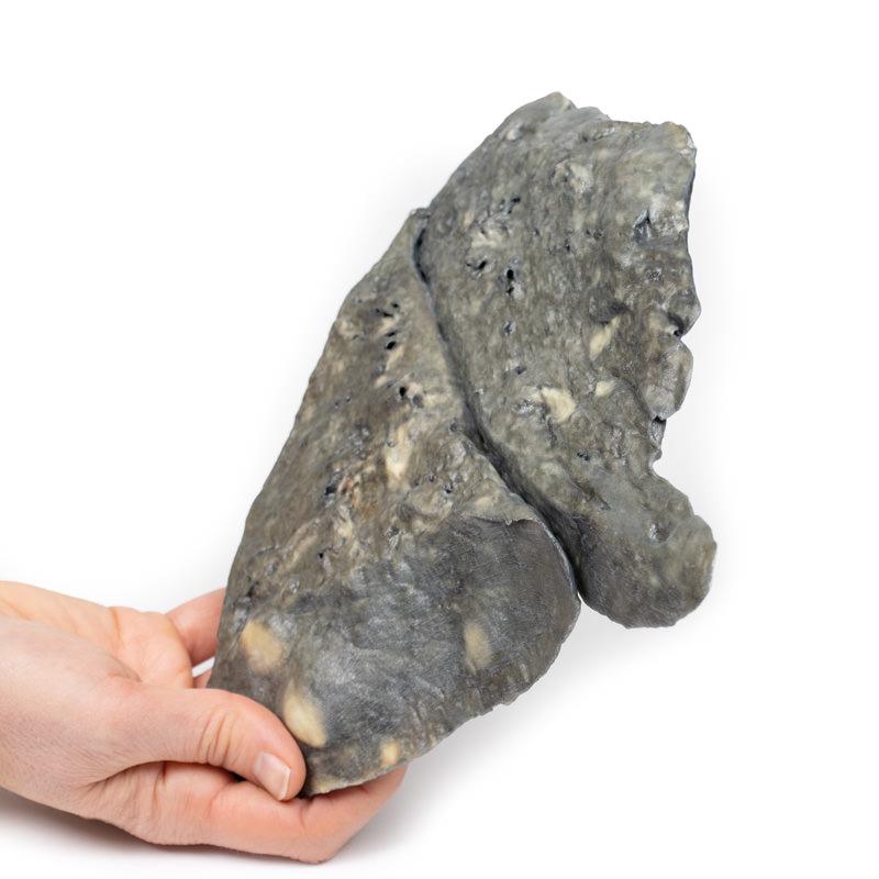
This model brings metastatic adenocarcinoma to life by highlighting tumor deposits scattered throughout lung tissue and along the pleural surfaces. Lung Multiple Secondary Carcinoma Deposits in The Lung and Pleura accurately showcases the distribution of nodules from 2 mm to 2 cm, the confluent masses near the hilum, and thickened pleura. Educational institutions, pathology labs, and medical professionals rely on its clinical accuracy for teaching metastatic disease mechanisms, diagnosis, and progression in a visually compelling way.
Features and Benefits
- 3D printed clinical-grade replica based on real pathological data
- Shows multiple pale tumor nodules throughout lung substance
- Highlights confluent nodules near the hilum and involvement of hilar lymph nodes
- Displays nodules beneath thickened costal, mediastinal, and diaphragmatic pleura
- Illustrates secondary metastatic adenocarcinoma deposits
- Enables tactile, visual learning and review of complex metastasis
- Durable and easy to handle for repeated educational use
Indications for Use
- Medical school and anatomy teaching involving pulmonary metastasis
- Pathology education about metastatic spread in lungs and pleura
- Professional development for clinicians and healthcare trainees
- Patient education regarding secondary cancer progression
Size Guide
- Full-scale model representing adult left lung anatomy
- Pale tumor nodules range in size from 2 mm to 2 cm
- Includes thickened pleural sections, hilar lymph nodes, and all described anatomical landmarks