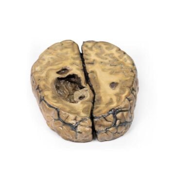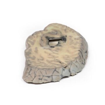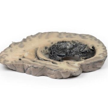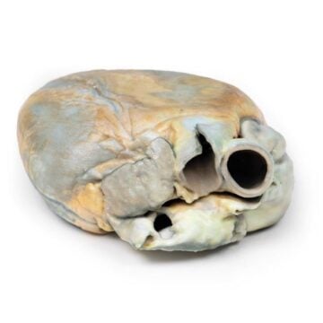

Features
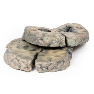
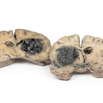
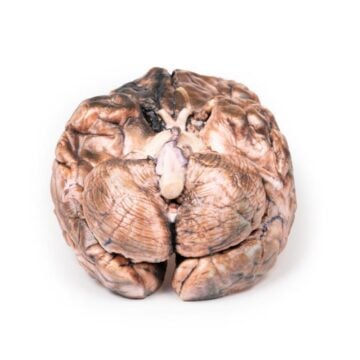
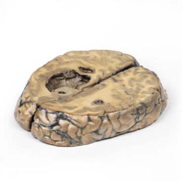
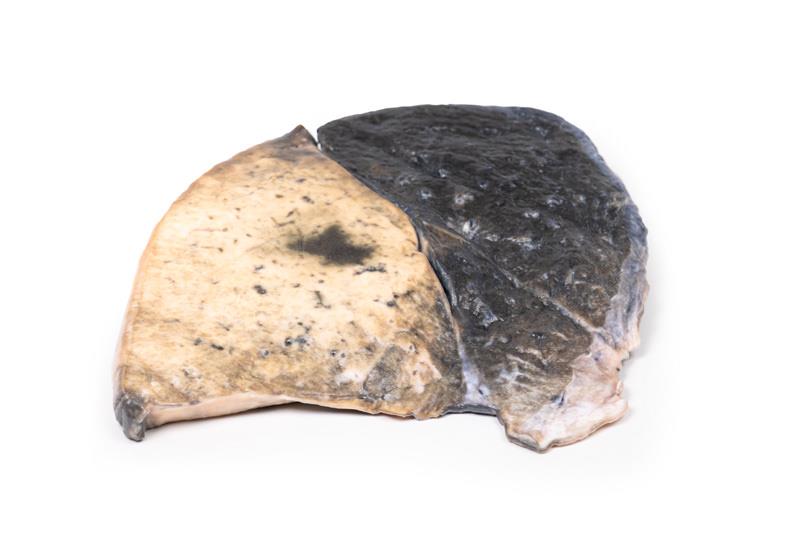
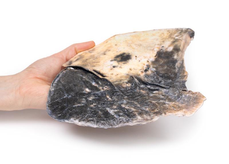
The Lobar pneumonia is a high-resolution 3D printed model of a parasagittal section of the right lung, created for medical educators, clinicians, and students. It provides an accurate visual representation of lobar pneumonia pathology, enabling clear demonstration of disease processes and lung anatomy. This educational model helps you visualize and explain the appearance and clinical features of lobar pneumonia better than flat images or diagrams.
See Lung Pathology Like Never Before with the Lobar pneumonia 3D Model
This premium anatomical model highlights the distinct boundaries between the three right lung lobes. The upper and middle lobes display realistic congestion and hyperemia, showing the darker visuals seen in true clinical cases. Smalls foci in the left lung add extra detail for advanced study. The model demonstrates classic red hepatization and parenchymal consolidation found in lobar pneumonia, simulating the solidified tissue as seen in actual pathology. Created for use in classrooms, hospitals, and medical simulation, it supports hands-on learning about pneumonia’s anatomical and radiological presentation.
Features and Benefits
- 3D printed, clinical-grade quality for authentic anatomical detail
- Exact representation of a parasagittal section of the right lung
- Distinct boundaries between upper, middle, and lower lobes
- Highlights upper and middle lobe congestion and hyperemia
- Includes smaller foci in the left lung for comprehensive study
- Accurately shows red hepatization and consolidation of lung parenchyma
- Excellent for teaching inflammatory exudate and alveolar consolidation
- Durable and ideal for repeated educational use
Indications for Use
- Medical and nursing education
- Clinical teaching about lobar pneumonia
- Radiology and pathology demonstrations
- Simulation-based training in lung disease
- Patient and caregiver education regarding lung conditions
Size Guide
- Model SKU – ERLMP2057
- Replicates anatomy of adult right lung
- Clinical-grade 3D printed material
- Exact sectioning for optimal visibility
- Dimensions match standard parasagittal lung sections (specific measurements not provided in source material)