Features
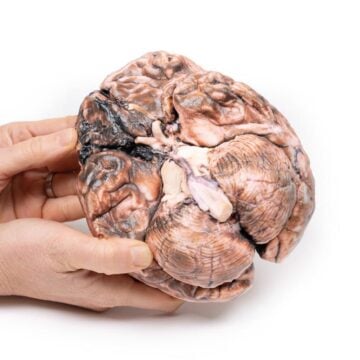
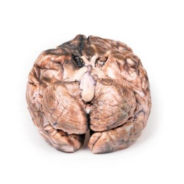
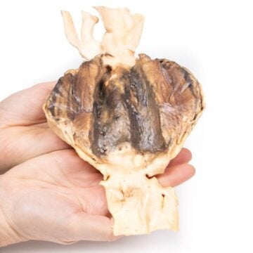
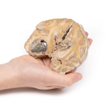
The Hepatic duct calculi and Obstructive Biliary Cirrhosis is a high-fidelity 3D printed anatomical model designed for medical educators and students. It demonstrates the impact of intrahepatic gallstones and obstructive biliary cirrhosis on liver structure. This model helps learners visualize and understand complex biliary pathology with exceptional clarity.
Bring Pathological Liver Anatomy to Life with Realistic 3D Visualization
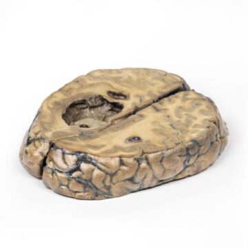
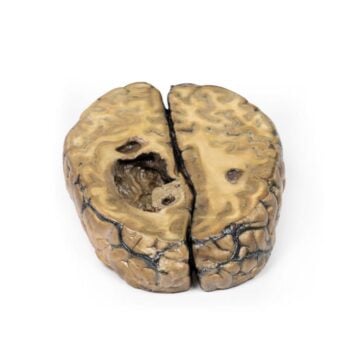
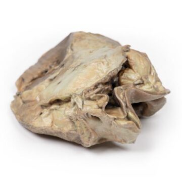
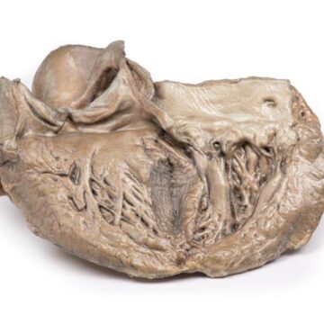
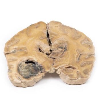
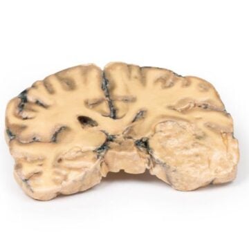
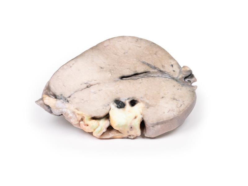
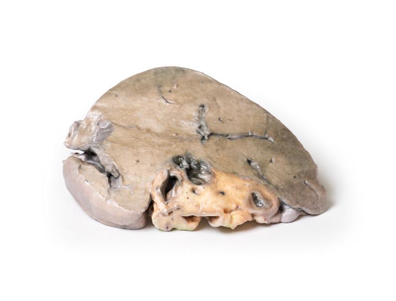
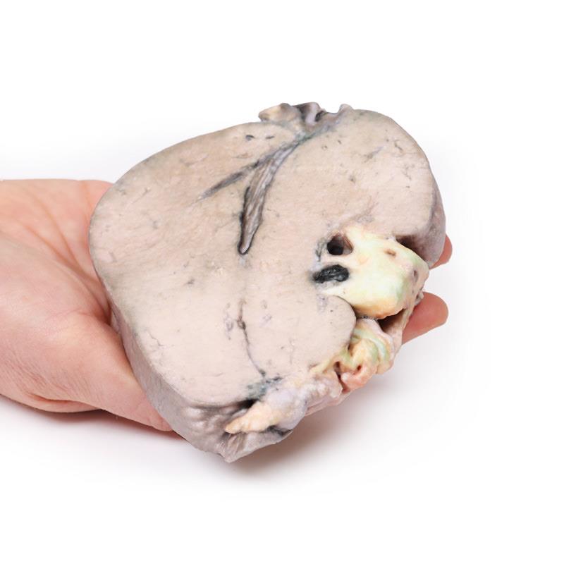
This model captures a liver slice with thickened capsule, finely nodular parenchyma, and dilated intrahepatic bile ducts. You will see a 10 mm pigmented calculus impacted in the hepatic duct and a 3 mm stone dislodged, representing secondary biliary cirrhosis caused by obstruction from calculi. Using the Hepatic duct calculi and Obstructive Biliary Cirrhosis model, educators can illustrate consequences of hepatolithiasis, including cholangitis, bile duct proliferation, and periportal fibrosis. Whether for pathology instruction or clinical training, this professional-grade model brings clarity to complex topics in liver disease.
Features and Benefits
- High-resolution 3D printed liver model with sectioned surface display
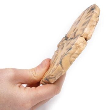
- Depicts thickened liver capsule and finely nodular structure
- Visible intrahepatic duct dilation with two pigmented gallstones (10 mm and 3 mm)
- Shows distended hepatic duct and features of secondary biliary cirrhosis
- Precisely highlights bile duct changes, periportal fibrosis, and inflammatory response
- Clinical-grade quality for medical education and demonstration
- Exceptional tool to explain hepatolithiasis, bile infarcts, and risk of cholangiocarcinoma
- Durable and easy to handle for repeated classroom use
Indications for Use
- Medical and anatomy education on liver pathology
- Pathology demonstration of intrahepatic gallstones and cirrhosis
- Simulation of secondary biliary cirrhosis features
- Clinical training in surgical, medical, and allied health settings
Size Guide
- Model displays a realistic liver slice with a 10 mm calculus and a 3 mm calculus
- Exact anatomical proportions and structures based on clinical specimens
- Overall model dimensions follow authentic hepatic morphology