Features
The HD Digihuman Virtual Anatomy Table is a premium digital teaching platform crafted for medical educators and students. It blends real human body data with advanced 3D reconstruction to offer a realistic virtual anatomy learning experience. Choose this system for its unrivaled clarity, ultra-high definition visuals, and fully interactive touch controls that bring human anatomy to life.
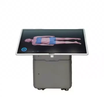
Revolutionize Medical Training with the HD Digihuman Virtual Anatomy Table
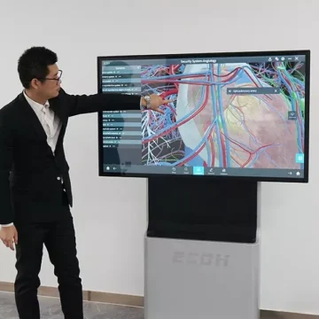
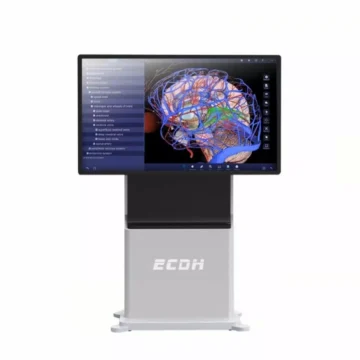
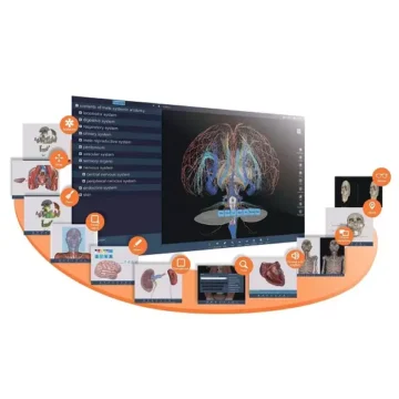
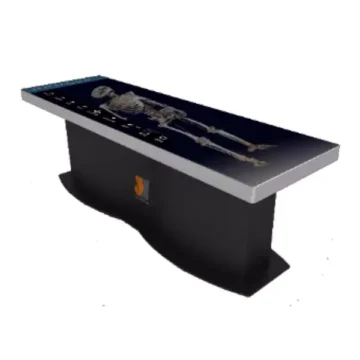
Transform anatomy education with this table, which lets you explore actual human body structures reconstructed layer by layer. The 88-inch UHD screen lifts and tilts for group or individual learning. Touch control grants total freedom to observe, rotate, and section human anatomy in any orientation or depth. The HD Digihuman Virtual Anatomy Table features high-resolution male and female data sets, over 3,000 3D structures, and instant access to clinical cases, slice libraries, and embryology modules. Advanced interactive functions support dynamic teaching, diagnosis practice, and precise anatomical study for accurate and engaging education.
Features and Benefits
- Real human body data with 0.1mm isotropic accuracy
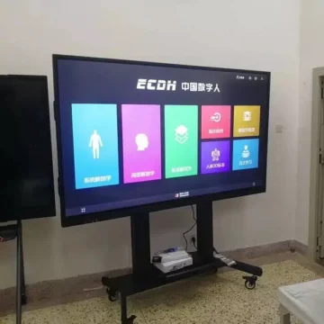
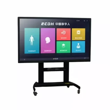
- 88-inch UHD touch screen with 3840 x 1080 resolution
- Screen lifts, lowers, and tilts to 90 degrees
- Over 3,000 3D anatomical structures and images
- Linear, curved, and orthogonal virtual cutting tools
- Interactive gesture controls for rotate, zoom, and pan
- Histology and pathology digital slice library
- Clinical cases with CT/MRI images and variable windowing
- Marking, dyeing, measuring, and annotation tools
- Individual multi-user accounts and custom presets
- High-performance specs – i7 10th Gen+, 64GB RAM, RTX 3080 graphics, 2TB SSD
- CE and FCC certifications
Indications for Use
- Medical education and training for students and professionals
- Teaching detailed human anatomy and sectional anatomy
- Simulation of clinical diagnoses using real case data
- Hands-on interactive anatomy in group or solo settings
Size Guide
- Screen size – 88 inches
- Screen resolution – 3840 x 1080
- Brightness – 700 cd/m²
- Contrast ratio – 1100 -1
- Vertical lift and 90-degree tilt capability