

Features
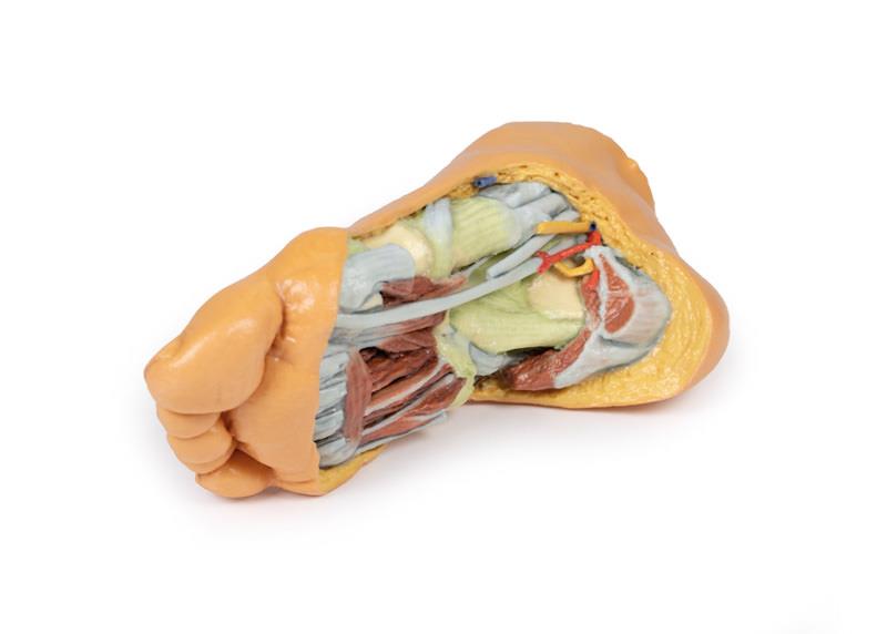
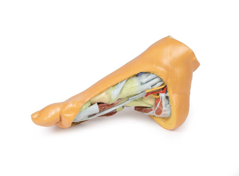
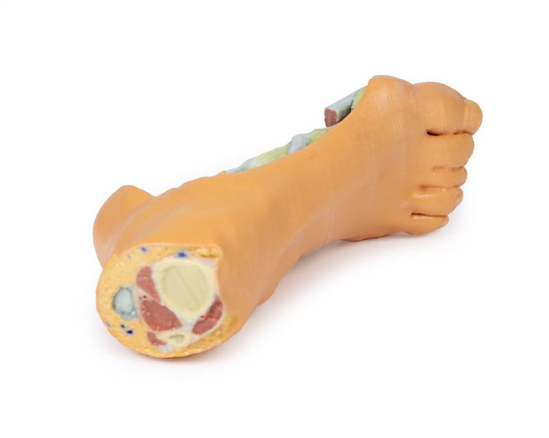
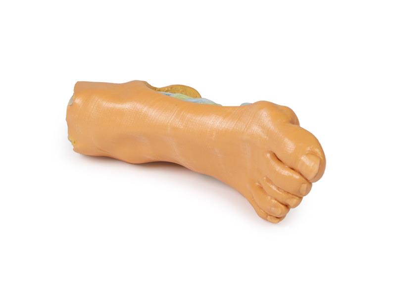
The Foot – Deep plantar structures is a 3D printed right foot anatomical model designed for educators, clinicians, and students. It provides a precise view of the deep plantar structures, making complex anatomy accessible and practical for teaching and demonstration. Choose this model for unparalleled anatomical accuracy and ethical simplicity over traditional specimen handling.
Reveal Exceptional Anatomical Detail with the Foot – Deep plantar structures
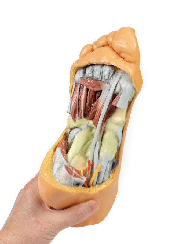
This clinical-grade 3D model unveils the intricate layers of the right foot’s plantar region. Expertly fabricated, it highlights the cut edge of the great saphenous vein within the superficial fascia, placed anterior to the medial and lateral plantar arteries and nerves. The design eliminates superficial fascias and aponeurosis, fully exposing the third muscular layer for deep structure study. Cut edges of the first, second, and third muscle layers remain for orientation, alongside the tendons of the flexor digitorum longus and brevis. Key muscular structures such as the adductor hallucis, flexor hallucis longus and brevis, as well as plantar interossei are visible. Exposed tarsal and metatarsal ligaments and the lateral aspect detailing peroneal tendon insertions enrich anatomical understanding. Benefit from ethical sourcing, unrivaled accuracy, easy storage, cost savings, and customizability, all in one model.
Features and Benefits
- 3D printed from real human anatomy for maximum precision
- Deep plantar structures of the right foot clearly exposed
- Medial view shows great saphenous vein and major plantar arteries and nerves
- Superficial fascia, plantar aponeurosis, and superficial musculature removed for clear visualization
- First, second, and third muscular layers retained at the calcaneus for orientation
- Flexor digitorum longus and brevis tendons featured
- Adductor hallucis heads visible beneath flexor hallucis longus tendon
- Plantar interossei accessible for detailed study
- Reveals tarsal and metatarsal joint capsule ligaments, long and short plantar ligaments, and the plantar calcaneonavicular ligament
- Lateral section reveals peroneus longus and brevis tendon insertions
- Printed to order for bespoke anatomical accuracy
- Outperforms cadaveric specimens for consistency, safety, and ease of use
- Allows customization for educational needs, including structure highlighting and cutaways
Indications for Use
- Medical, podiatry, and physiotherapy education
- Clinical demonstration of plantar foot anatomy
- Patient education regarding foot structure and pathology
- Anatomical study and surgical planning
- Standardized teaching and examination
Size Guide
- Life-size model replicates actual anatomical dimensions of an adult right foot
- Model SKU – ERLMP1940