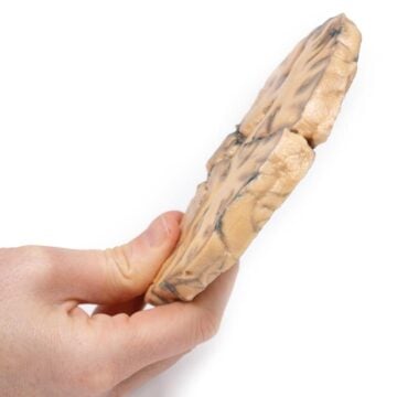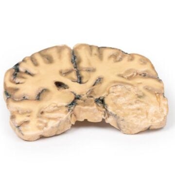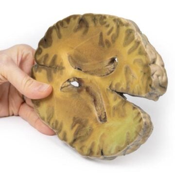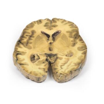
Features
The Chondrosarcoma of scapula is a clinical-grade 3D anatomical model designed for medical educators, students, and healthcare professionals. It helps users understand the pathology, clinical presentation, and structure of chondrosarcoma affecting the scapula. Choose this model for its unmatched educational detail, accuracy, and real-life replication of a rare bone tumor scenario.
Accurately Visualize Malignant Bone Tumour Pathology with This 3D Model
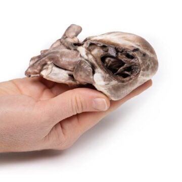
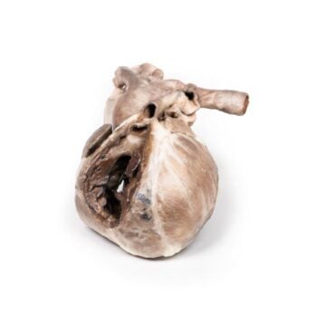
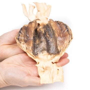
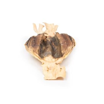
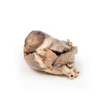
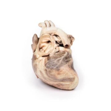
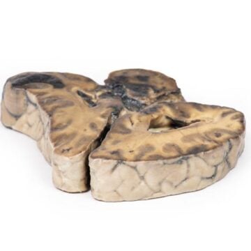
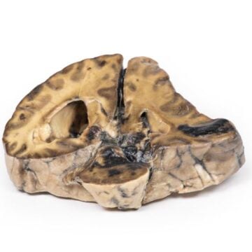
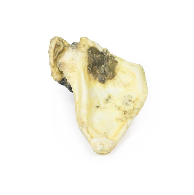
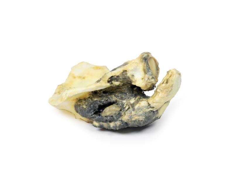
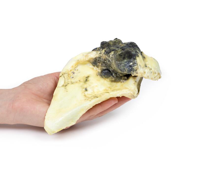
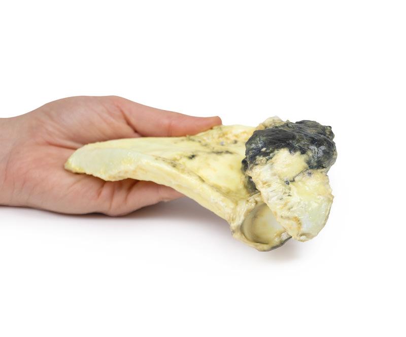
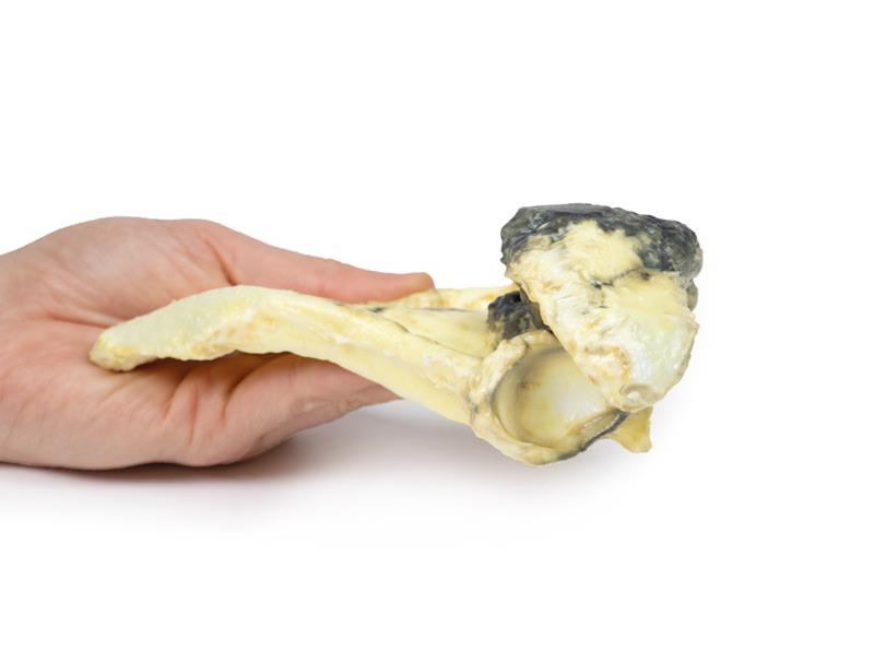
This unique 3D printed Chondrosarcoma of scapula model replicates a real patient specimen, capturing an irregular, lobulated tumour infiltrating the right scapula. The model features anatomical detail such as a mottled pale-yellow brown tumour with patchy haemorrhage, involvement of the acromion and coracoid process, and adherent muscle and fibrous tissue. The detailed anatomy supports hands-on learning across oncology, pathology, and orthopedic training. See key histological features including pleomorphic cells and cartilage formation, deepening your pathological understanding. Clinical-grade manufacturing ensures the accuracy necessary for professional education or research applications.
Features and Benefits
- Life-sized 3D printed model based on actual clinical case
- Replicates a lobulated tumour 11 cm in diameter
- Includes infiltration of the scapula, acromion, and coracoid process
- Shows mottled pale-yellow brown color and areas of surface haemorrhage
- Displays adherent muscle and fibrous tissue for realistic context
- Enables visualization of tumor replacing normal bone structure
- Delivers detailed histological characteristics for medical study
- Clinical-grade quality for teaching, exhibitions, and research
Indications for Use
- Medical education and anatomical demonstration of chondrosarcoma in the scapula
- Training in orthopedic and oncology case-based learning
- Pathology and tumor identification for clinicians and students
- Reference for surgical planning and clinical discussions
Size Guide
- Full right scapula with tumor inclusive
- Tumor maximum diameter measures 11 cm
- Replicates the original excised pathological specimen