
Features
The Chondrosarcoma of femur and ilium is a precision 3D printed model developed for educators, clinicians, and medical students. It provides an anatomically accurate representation of a rare, malignant bone tumor, supporting advanced pathology education and surgical planning. This model helps users visualize complex pelvic oncology cases, giving a clear advantage over 2D materials.
Explore Clinical Pathology with a Realistic 3D Tumor Model
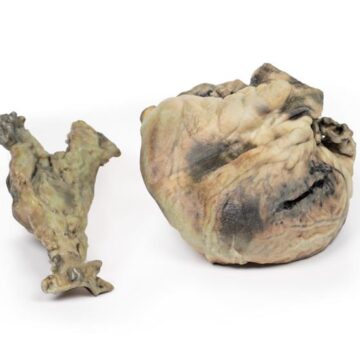
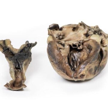
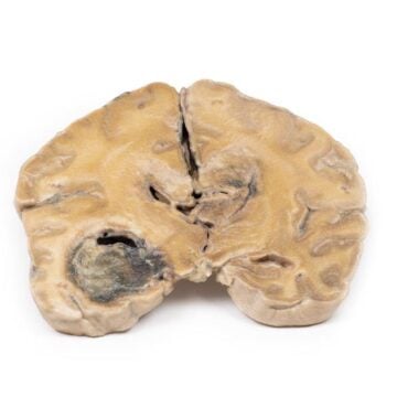
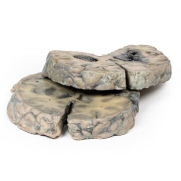
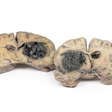
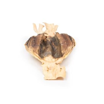
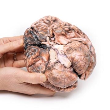
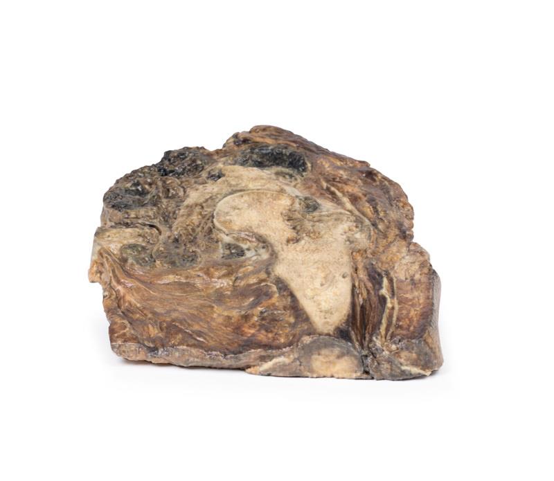
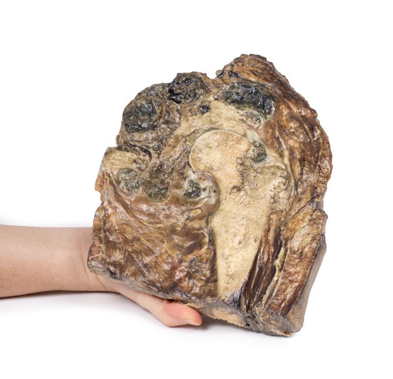
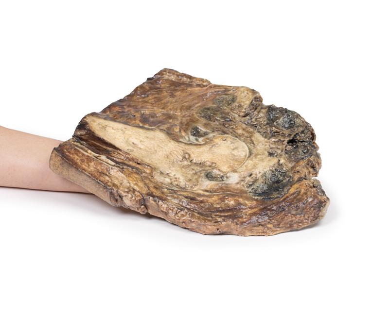
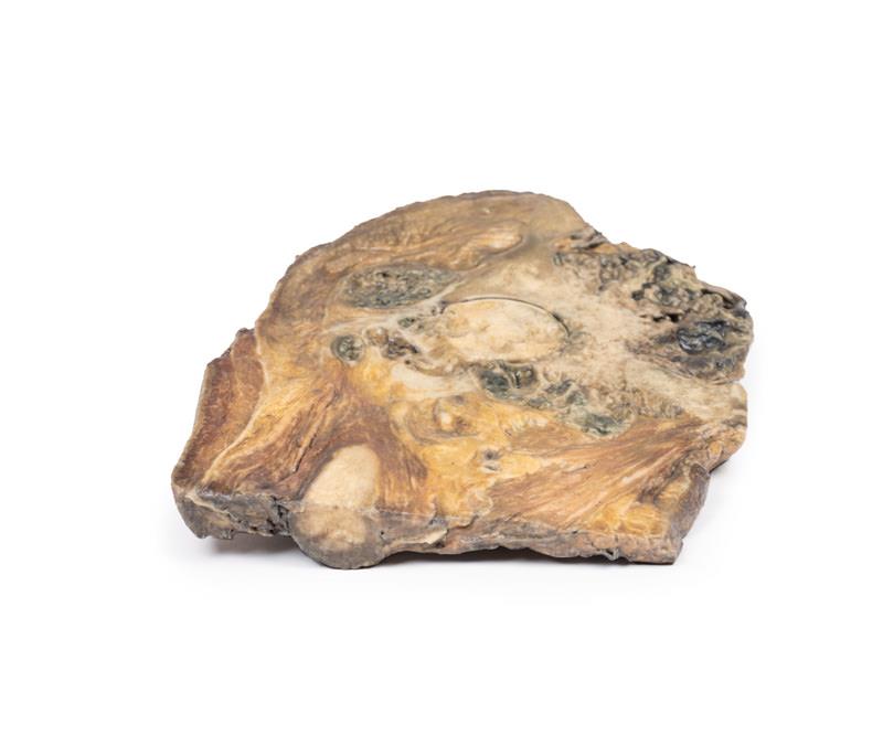
This Chondrosarcoma of femur and ilium model showcases the true anatomy and pathology of a lobulated, necrotic, and encapsulated malignant bone tumor, spanning the proximal femur into the ilium. With clinical-grade realism, users examine infiltration, cavitation, necrosis, and hemorrhage, facilitating discussion and practical demonstration. The model aids in understanding the technical challenges of pelvic tumor excision and reconstruction by accurately reflecting both soft tissue extension and clear anatomical boundaries. Developed to match clinical education standards, the model brings clarity to this complex and rare diagnosis, making it indispensable for specialized training sessions, demonstrations, and patient education.
Features and Benefits
- High-resolution 3D printed reproduction based on real pathological anatomy
- Demonstrates tumor extension from femur head and neck into ilium
- Displays macroscopic features of malignancy—cavitation, necrosis, hemorrhage, encapsulation
- Highlights infiltration into soft tissue beyond bone margins
- Enables hands-on teaching and pre-surgical planning
- Clinical-grade detail enhances educational value
- Durable construction for repeated academic use
Indications for Use
- Medical and surgical education on chondrosarcoma pathology
- Surgical planning for pelvic bone tumors
- Clinical demonstrations on tumor infiltration and resection margins
- Patient and trainee communication tool
Size Guide
- Full-scale anatomical reproduction—dimensions replicate the upper femur, femoral head and neck, and adjacent ilium
- Model dimensions vary based on patient anatomy, typically around 25-30 cm in length and 15-18 cm at its widest point
- No size code options—model provides true-to-life proportions for demonstration