
Features
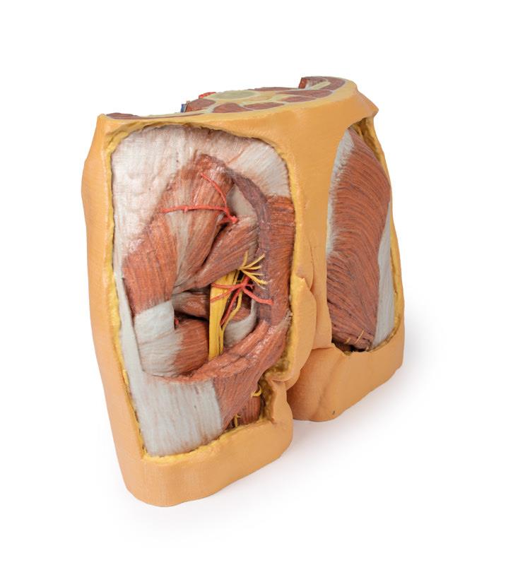
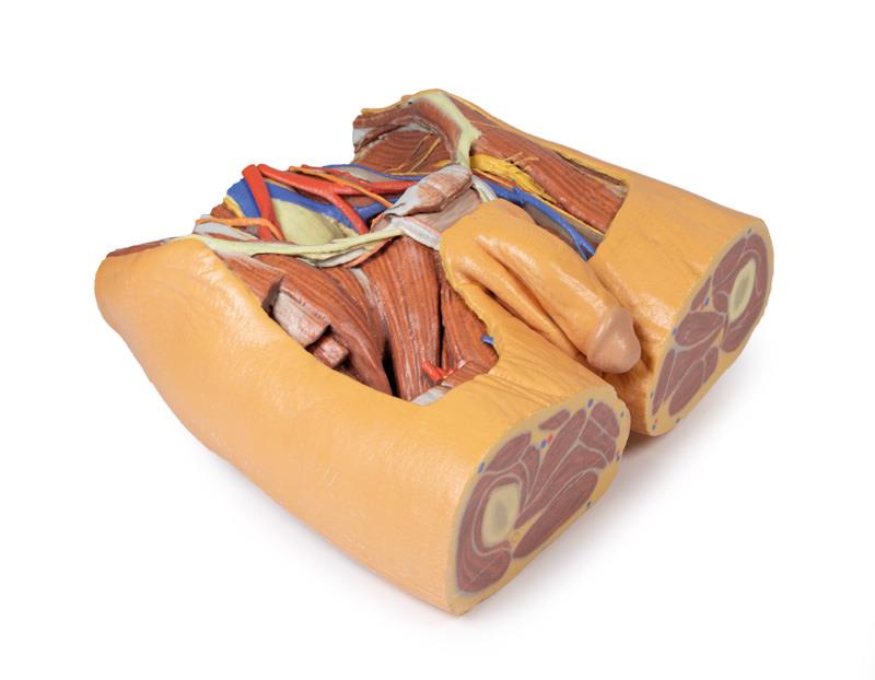
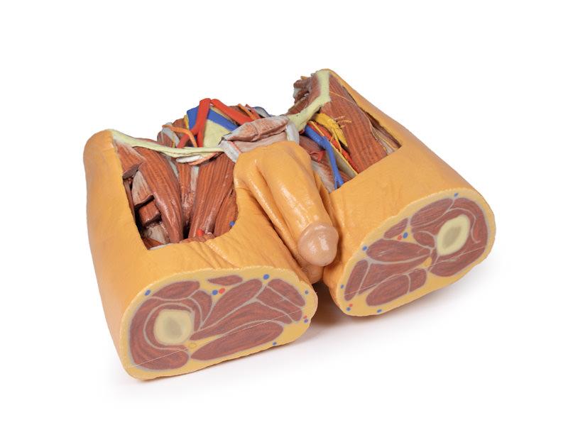
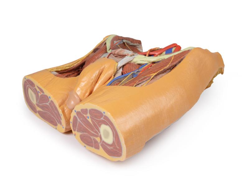
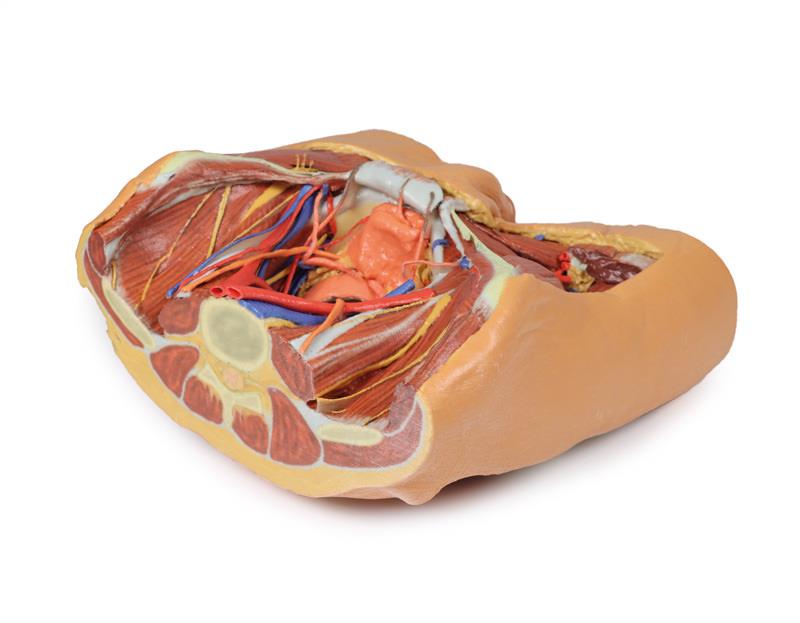
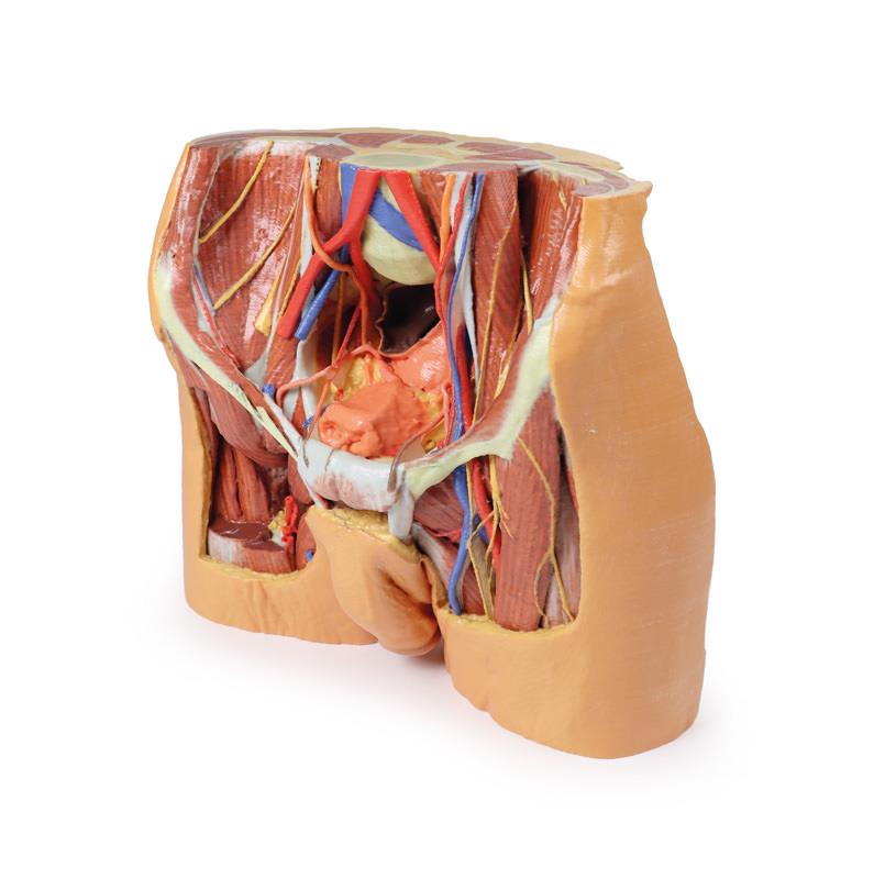
The Male Pelvis is a multipart 3D printed anatomical specimen designed for medical professionals, educators, and anatomy students. It provides a detailed, clinically accurate view of the male pelvic cavity, the lower posterior abdominal wall, and proximal thigh. Choose this product for its superior anatomical accuracy and unmatched teaching value.
Transform Learning and Clinical Demonstrations with the Male Pelvis 3D Model
This advanced 3D printed Male Pelvis model showcases remarkable realism and detail, allowing you to explore intricate pelvic structures with clarity. The specimen includes the inferior portions of the posterior abdominal wall, the pelvic cavity, and the upper proximal thigh, covering essential features like the gluteal region and femoral triangles. You can visibly trace the course of major vessels and nerves, including the common iliac veins forming the inferior vena cava, and the convergence of common iliac arteries. Detailed muscular structures such as the iliacus and psoas muscles are easy to identify, as are the branching nerves including ilioinguinal, lateral cutaneous nerve of thigh, femoral, and genitofemoral nerves. Important clinical details like the course of ureters and vas deferens are also clear, supporting both surgical training and anatomical education.
Features and Benefits
- Multipart, clinical-grade 3D printed model ensures exceptional anatomical accuracy
- Displays pelvic cavity, lower posterior abdominal wall, and proximal thigh in one specimen
- Covers gluteal regions and femoral triangles for comprehensive learning
- Clearly presents major arteries, veins, nerves, and ligaments
- Provides visible detail of iliacus, psoas muscles, and a prominent psoas minor tendon
- Shows the course of ureters and vas deferens
- High-quality print suited for repeated educational or clinical demonstrations
- Ships fast across Australia with tracked nationwide delivery
Indications for Use
- Anatomy education for students and faculty
- Surgical training and demonstration
- Patient education sessions
- Advanced clinical examination and reference
- Medical research and study
Size Guide
- Single multipart specimen—derived from a transection at approximately the L2/L3 intervertebral disc
- Displays anatomical structures from lower posterior abdominal wall to proximal thigh
- Exact anatomical sizing, matching realistic male pelvic dimensions for detailed study