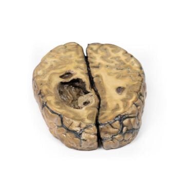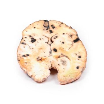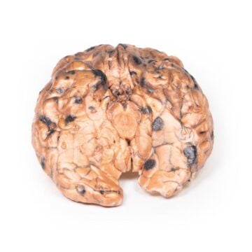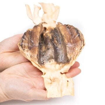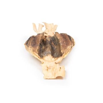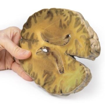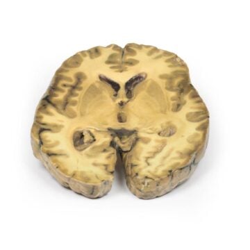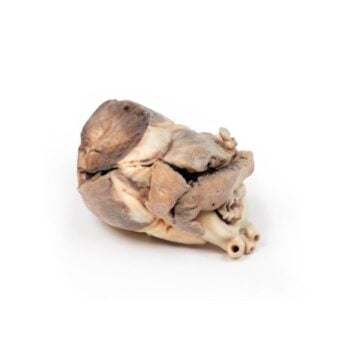
Features
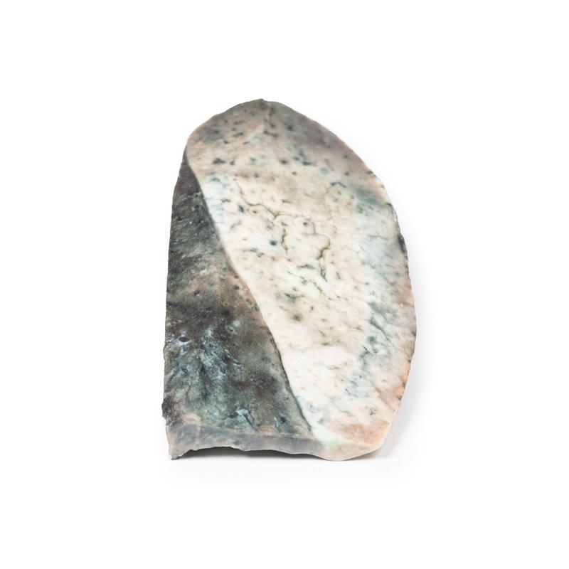
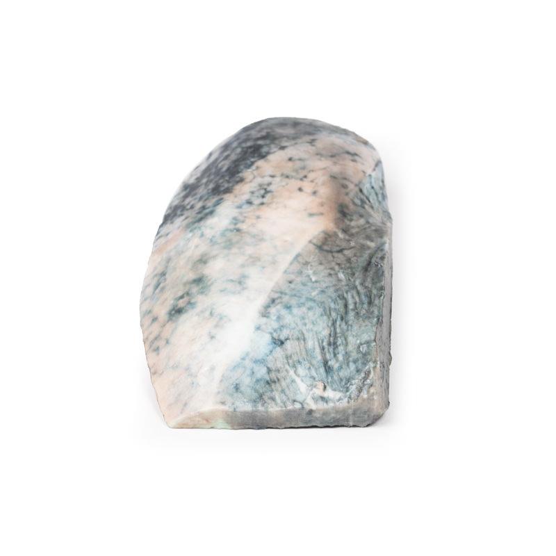
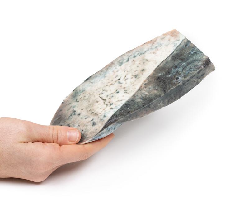
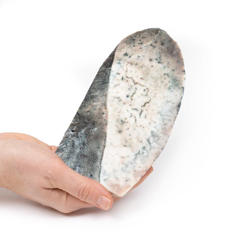
The Lobar pneumonia – Grey Hepatisation Phase is a clinical-grade 3D printed lung specimen designed for medical educators, students, and health professionals. It clearly demonstrates the late consolidation stage of lobar pneumonia, providing a realistic resource for anatomical and pathological study. Choose this model for its unparalleled accuracy and detail, elevating your anatomical understanding and teaching sessions.
Explore the Essential Pathology with Lobar pneumonia – Grey Hepatisation Phase
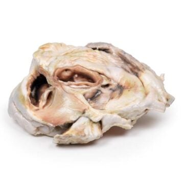
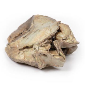
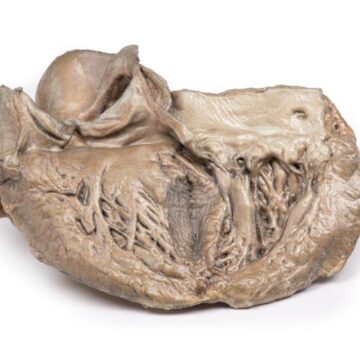
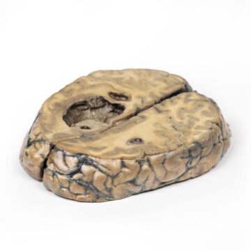
This 3D model captures a parasagittal section of the right lung, with distinct boundaries between the upper and lower lobes. The upper lobe appears pale grey and congested, mirroring the classic features of the grey hepatisation phase in lobar pneumonia. The model vividly illustrates consolidation caused by intra-alveolar inflammatory exudates, offering an immersive experience for learners. Designed to clinical standards, this replica provides insight into the progression of pneumonia, including macrophage infiltration and tissue restoration processes. Enhance your courses or clinical training with a hands-on educational tool that brings real pathology into the classroom or lab.
Features and Benefits
- Clinical-grade 3D printed model of the right lung
- Shows distinct upper and lower lobe boundaries
- Replicates grey hepatisation phase with pale, congested upper lobe
- Highlights consolidation through fibrinopurulent exudate
- Depicts macrophage and siderophage infiltration
- Demonstrates the process of pulmonary restoration after inflammation
- Provides excellent visual aid for medical education
- Durable and suitable for repeated classroom use
Indications for Use
- Medical and anatomy education
- Pathology demonstrations and lectures
- Clinical skills and training workshops
- Student laboratory studies
Size Guide
- One standard size model – represents a parasagittal section of an adult right lung with clear lobe demarcation