Features
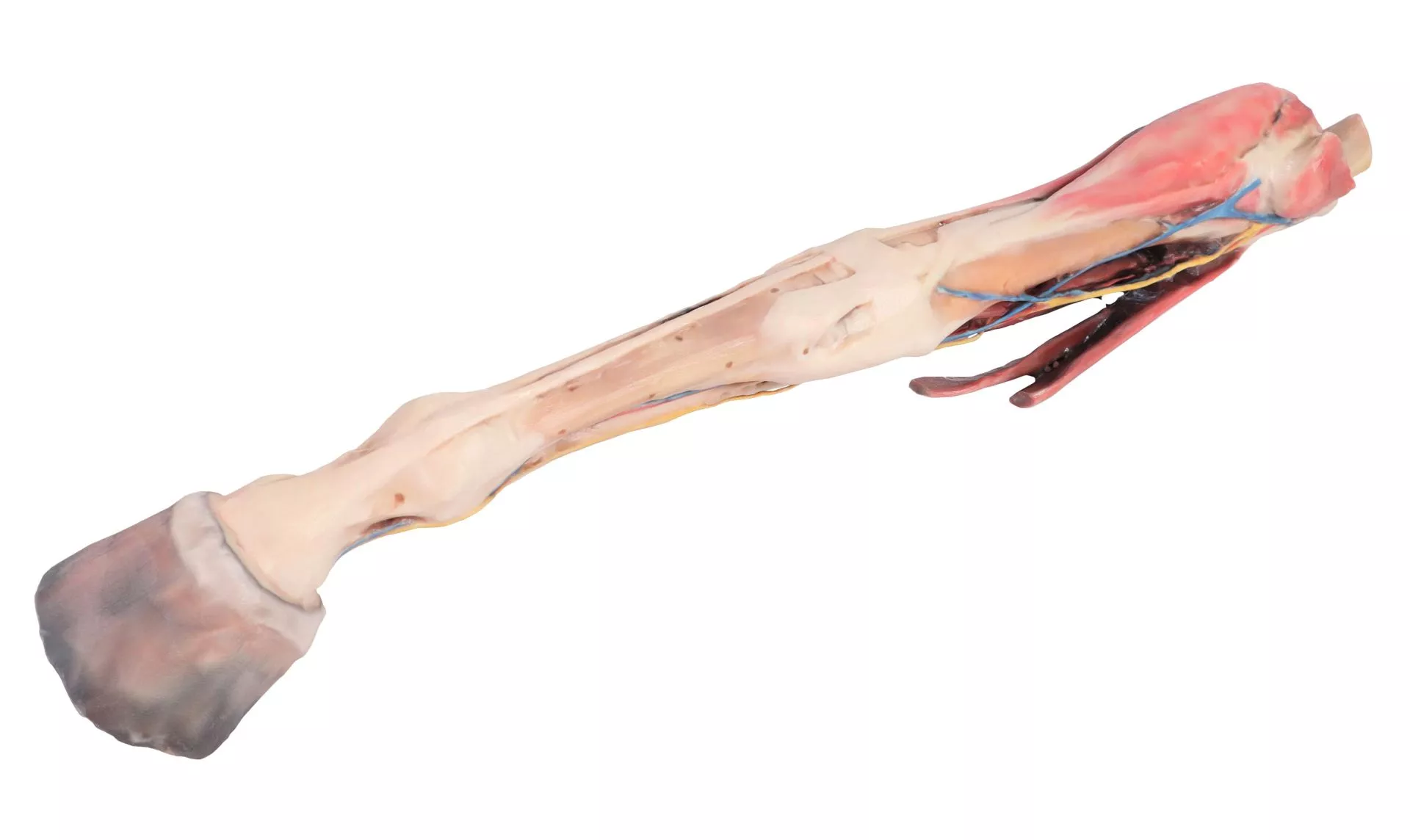
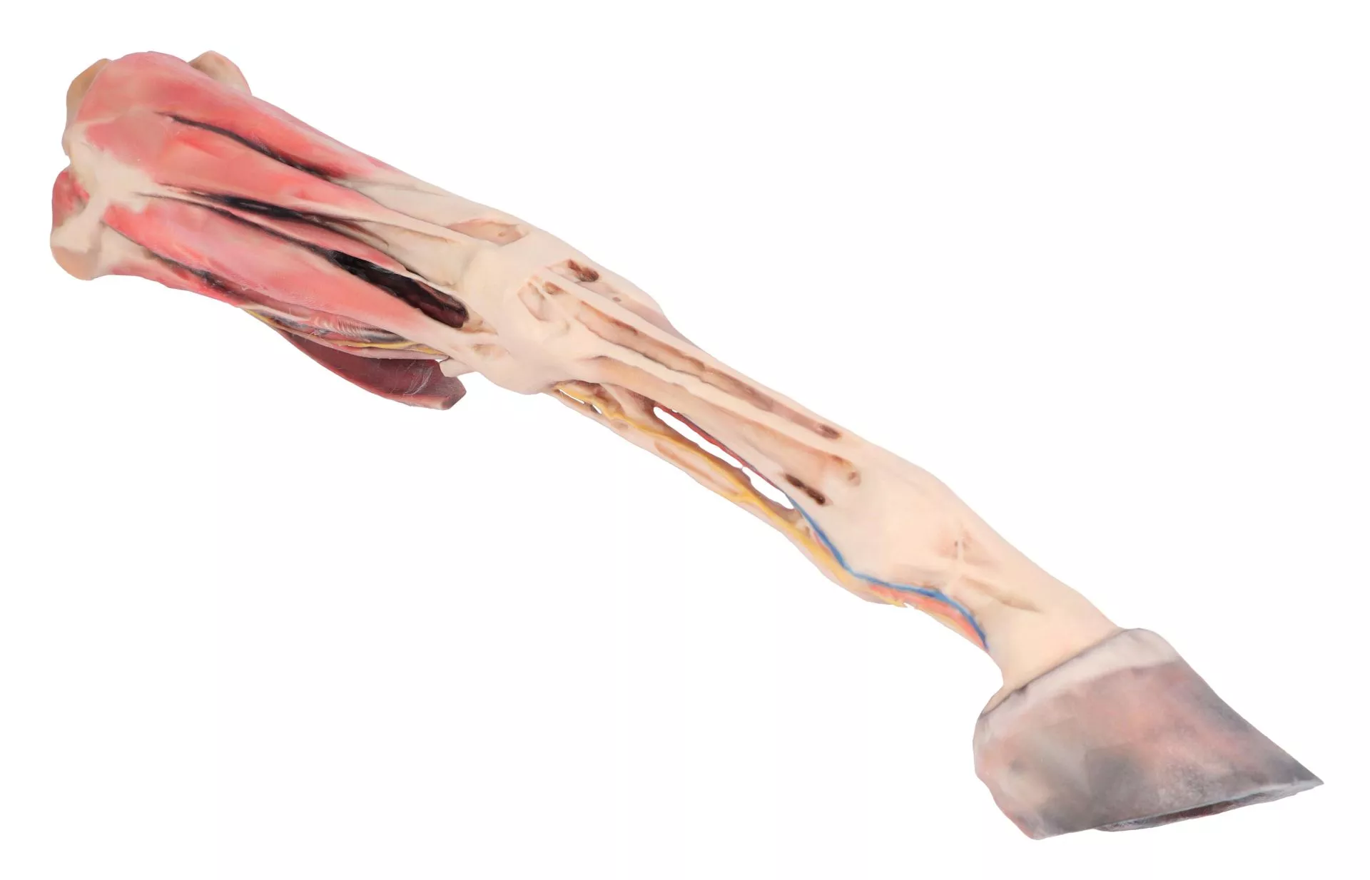
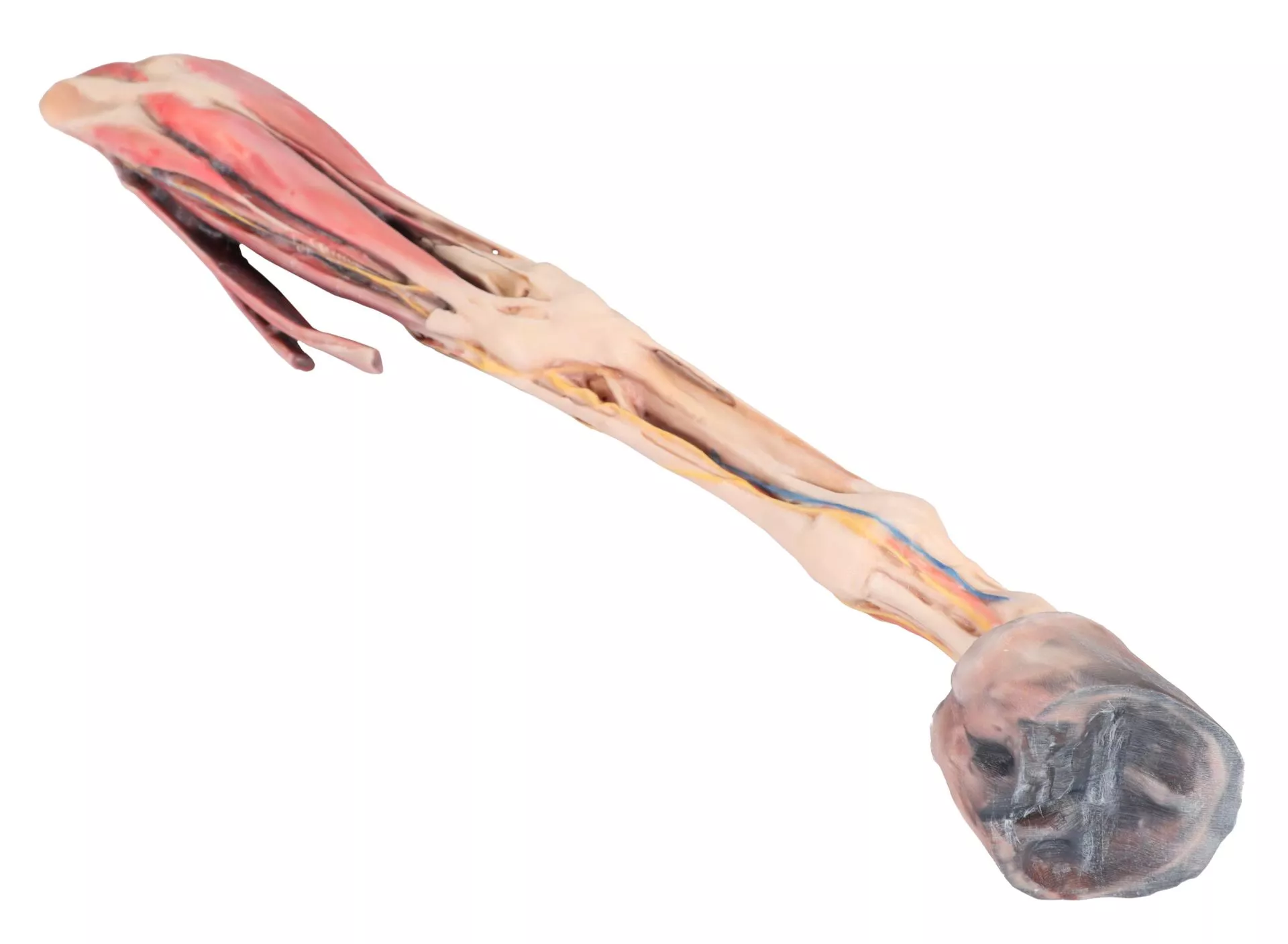
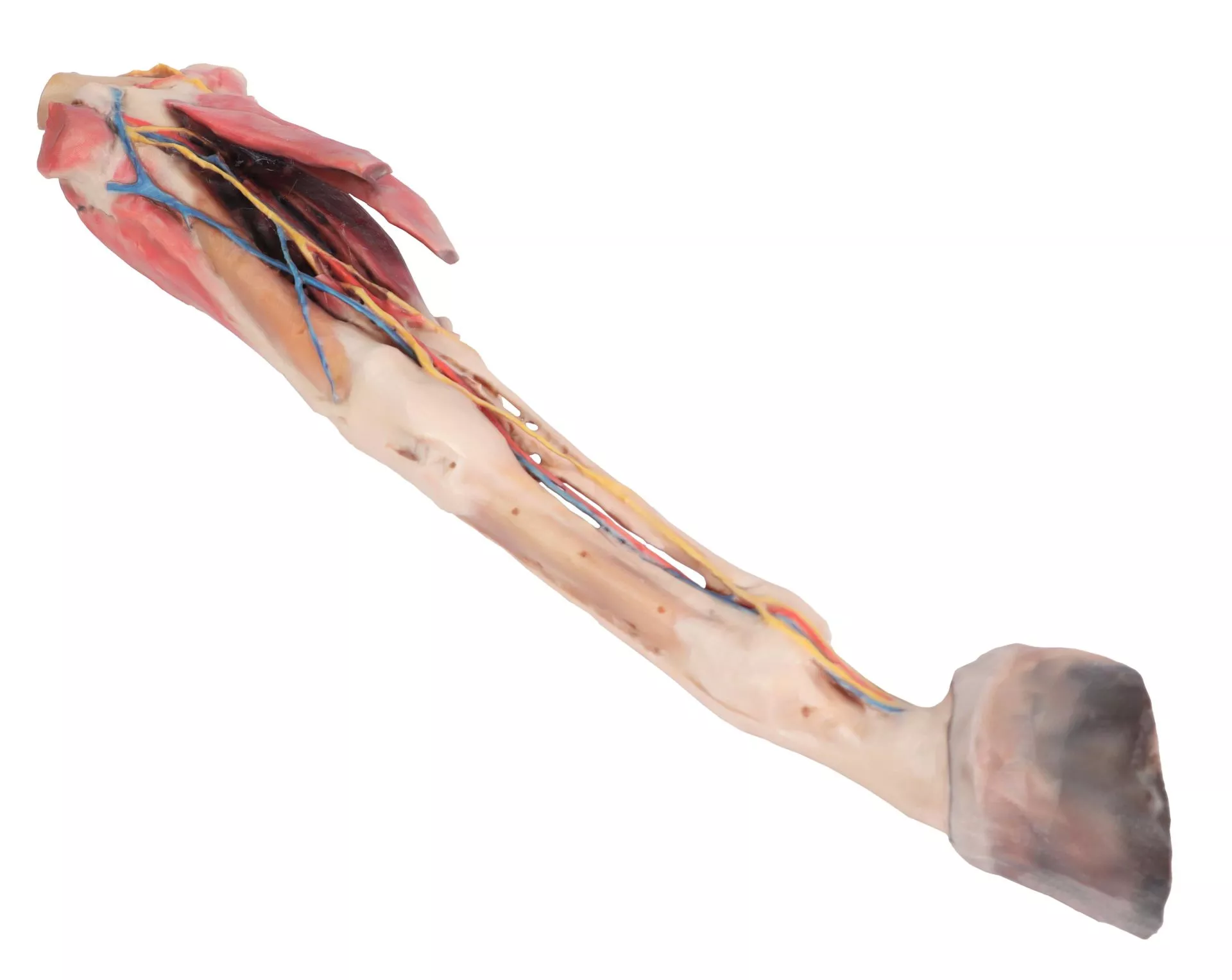
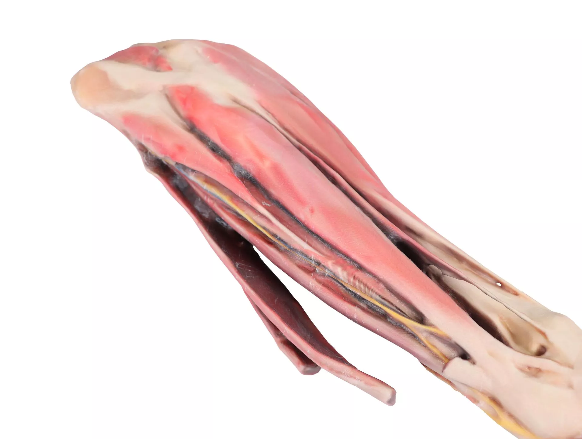
The Horse forelimb – muscles, tendons, ligaments vessels and nerves distal to the stifle – 1/3 life-size is a precise anatomical model created for veterinary professionals, educators, and students. It provides a clear, in-depth view of the equine forelimb’s anatomy, making it essential for teaching and clinical demonstration. Detailed preservation of structures ensures reliable visual reference and hands-on learning.
Unlock the Complex Anatomy of the Equine Forelimb
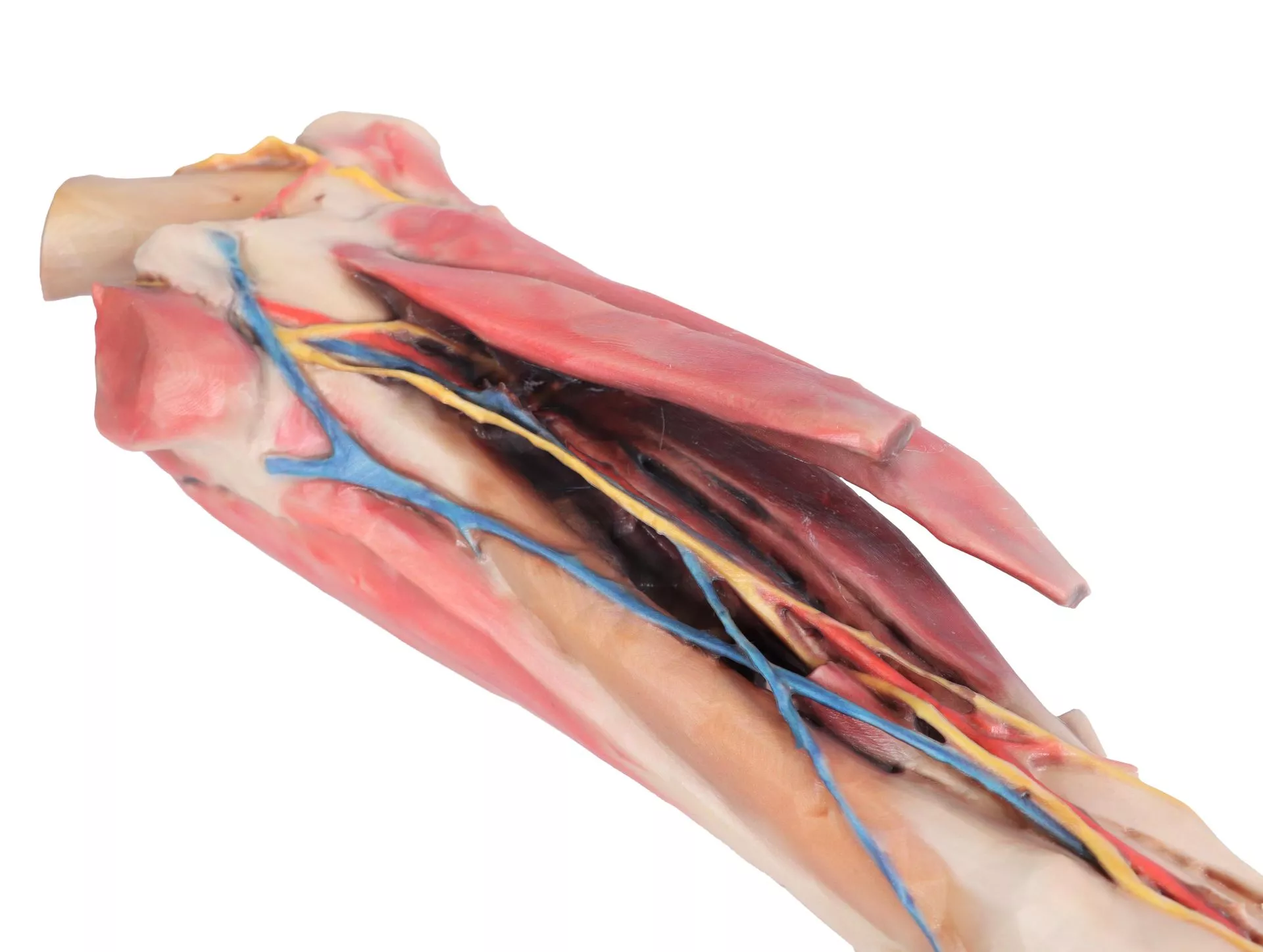
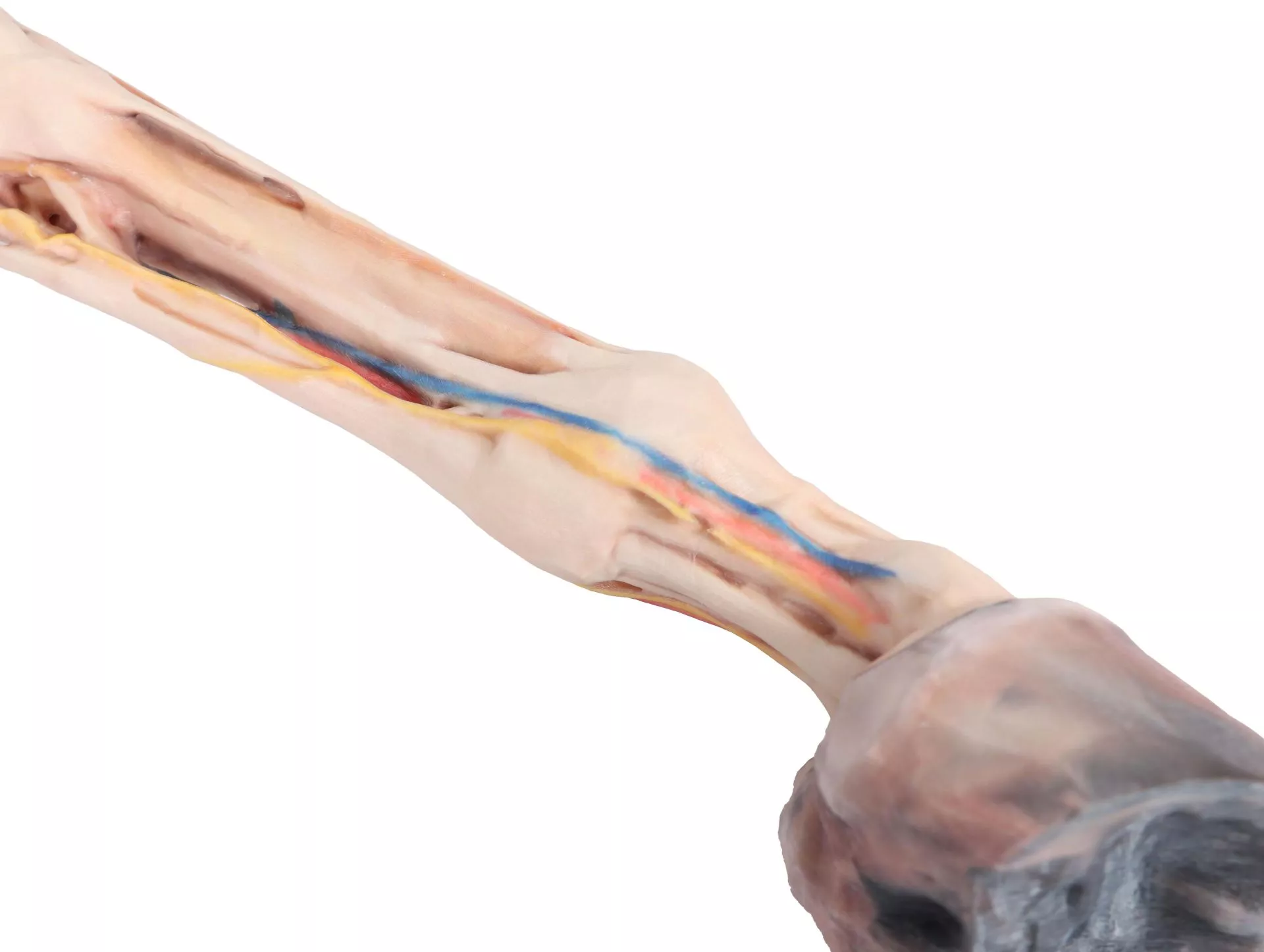
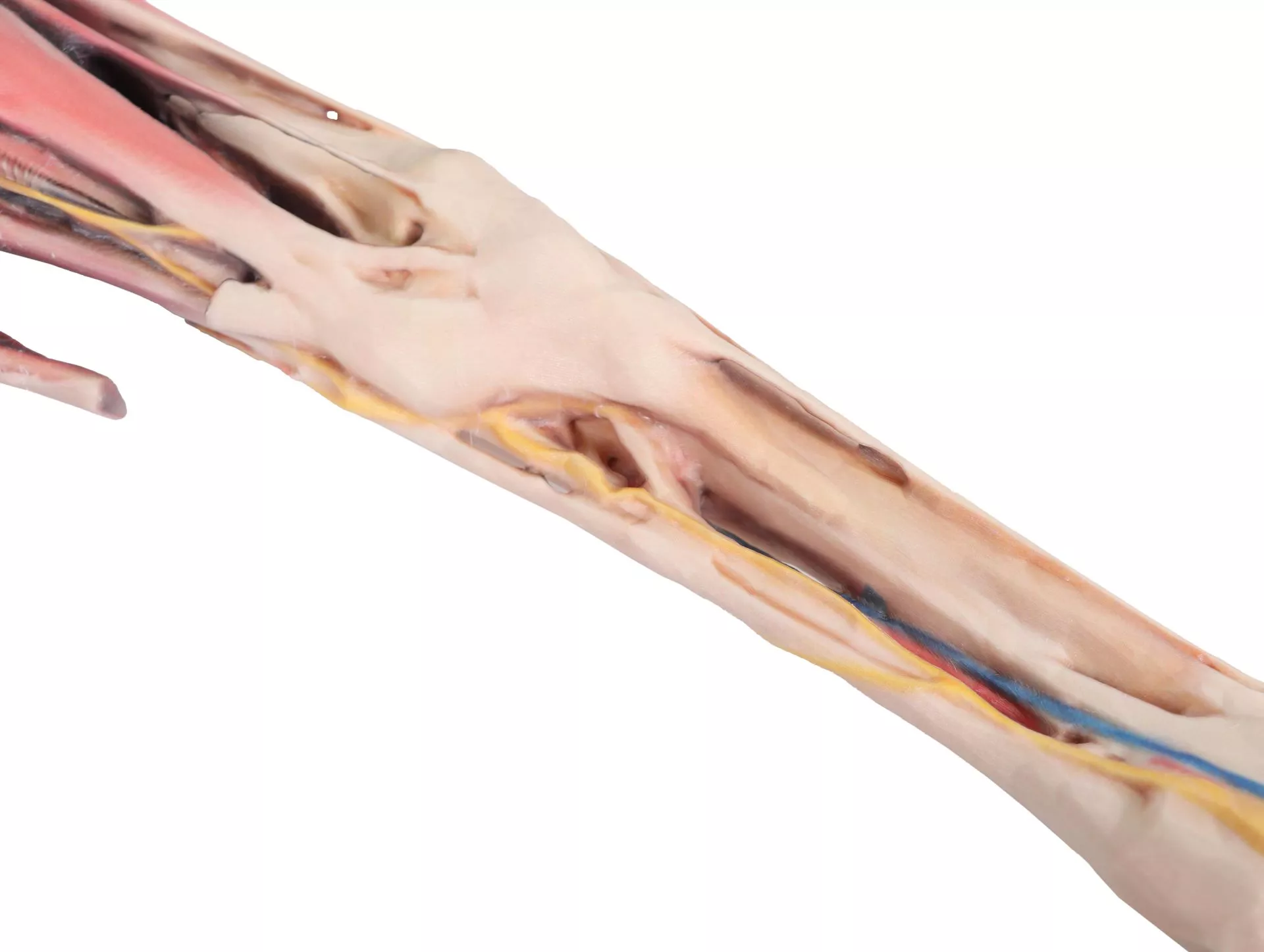
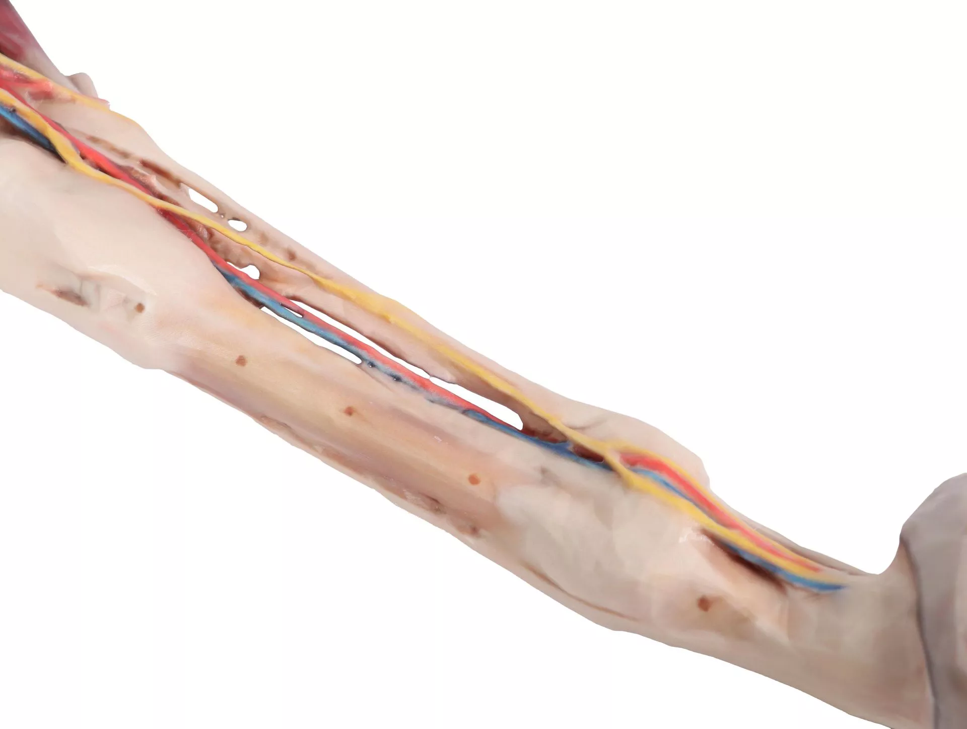
Gain comprehensive insight into equine anatomy with the Horse forelimb – muscles, tendons, ligaments vessels and nerves distal to the stifle – 1/3 life-size. This clinical-grade model meticulously presents the superficial anatomy of a right horse forelimb from the elbow to the hoof. Examine the pathways of the median and cubital nerves, alongside the extensor and flexor muscles of the carpus and digits. Major vascular structures remain visible, including the main branches of the median artery plus the median and cephalic veins. Palmar nerves are clearly delineated in relation to the digital tendon flexors, and detailed palmar digital neurovascular bundles are shown distal to the metacarpophalangeal joint. This model powers advanced learning, anatomical accuracy, and practical confidence.
Features and Benefits
- Displays the superficial anatomy of a right equine forelimb from elbow to hoof
- Shows carpal and digital extensor and flexor muscles with accurate anatomical placement
- Presents pathways of the median and cubital nerves
- Illustrates main branches of the median artery
- Includes preserved median and cephalic veins
- Shows topographic relation of palmar nerves with digital tendon flexors
- Reveals clear palmar digital neurovascular bundles beyond the metacarpophalangeal joint
- Constructed to 1/3 life size for clear visualization and space efficiency
- Clinical-grade quality ensures high educational value and durability
Indications for Use
- Veterinary anatomical education and demonstration
- Medical student and veterinary training
- Classroom and clinical instruction of equine anatomy
- Visual reference for research and clinical planning
Size Guide
- Scale is 1/3 of actual equine forelimb size
- Representative of superficial structures from elbow to hoof