Features
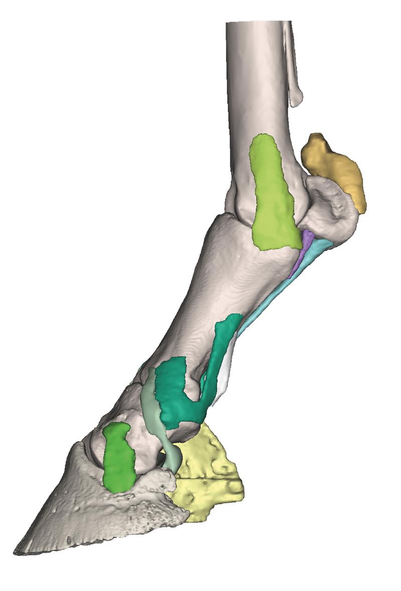
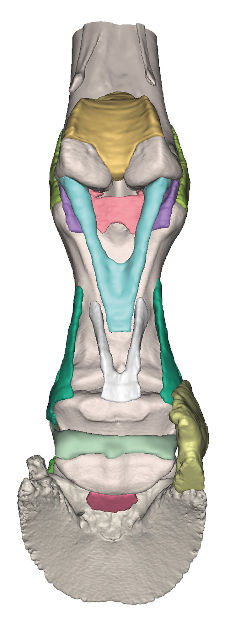
The Foot of a horse as model – Model 4 is a life-size 3D equine foot model designed for veterinary educators, professionals, and students. It reveals only the bones and deep supporting ligaments, allowing in-depth study of advanced anatomical structures. Choose this detailed model to gain a clear, hands-on understanding of the horse’s foot beyond what standard diagrams can offer.
Study Equine Limb Structure with Unmatched Detail Using the Foot of a horse as model – Model 4
Step into advanced anatomical education with a model crafted from real clinical CT and MR scans. Every bone and ligament appears true to life, 3D printed in full color with precise, individually colored structures. By removing the tendons and suspensory ligament, this model focuses attention on the core support system of the distal limb. The hoof capsule is available separately and anchors the limb in a natural stance, making this model essential for expert demonstrations or lessons. Perfect for in-depth veterinary learning, this model belongs to a series, with Model 4 representing the most reduced anatomical view for advanced exploration.
Features and Benefits
- Derived from CT and MR co-registered data for clinical accuracy
- Life-size 3D printed replica
- Each anatomical component is individually colored for clarity
- Shows bones and deep supporting ligaments with tendons and suspensory ligament removed
- Full-color printing enhances anatomical differentiation
- Hoof capsule available separately for stance positioning
- Durable education-standard quality construction
Indications for Use
- Veterinary student anatomy education
- Professional demonstration in clinical or teaching settings
- Study of equine musculoskeletal structures and pathology
- Reference for equine podiatry and advanced limb dissection studies
Size Guide
- Life-size equine distal limb representation
- Modeled after actual horse CT and MR scan data for anatomical precision