




Features
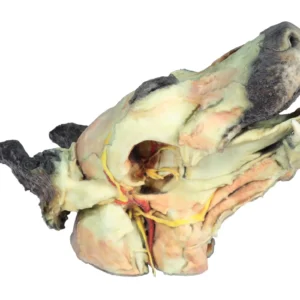
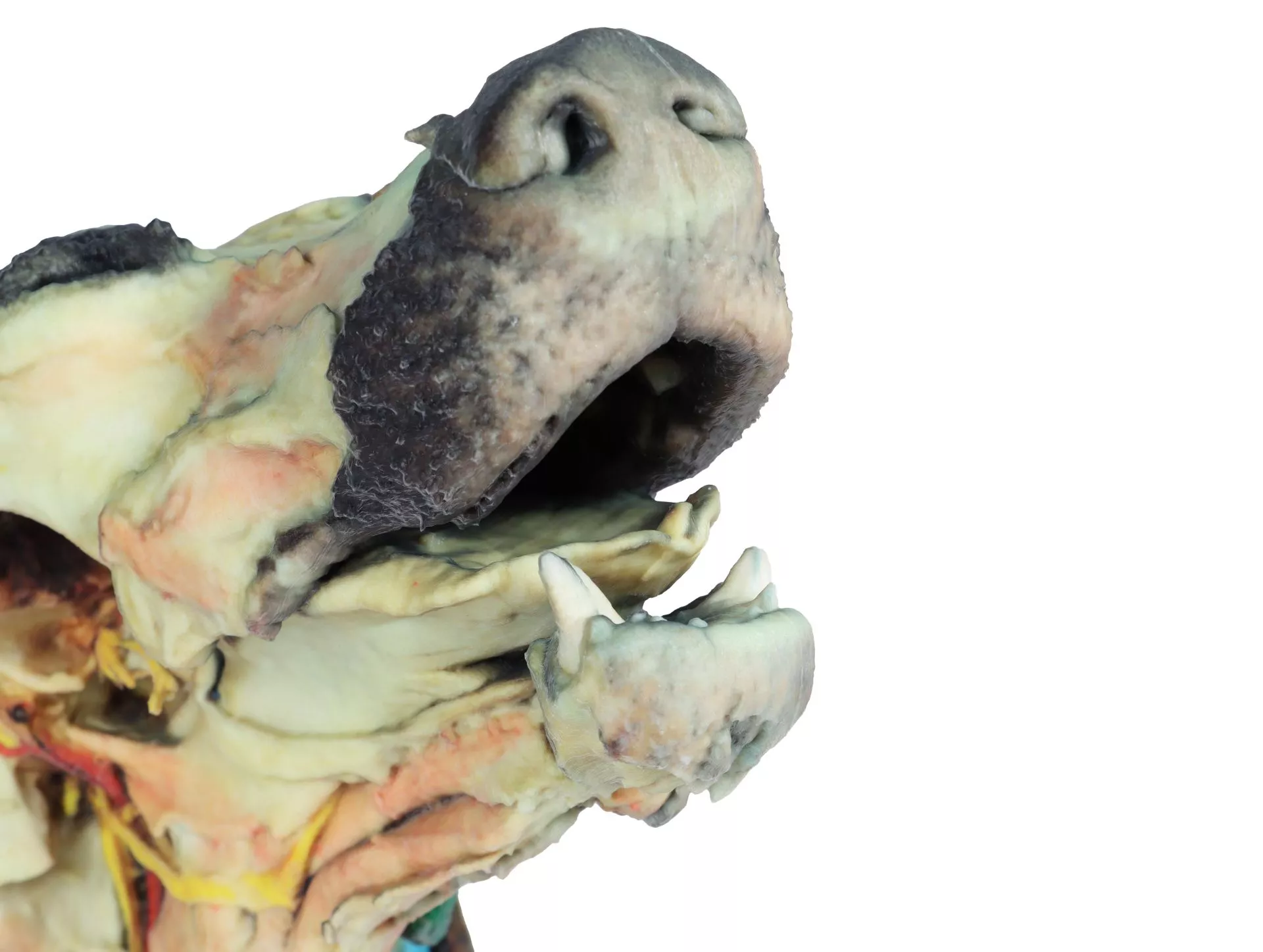
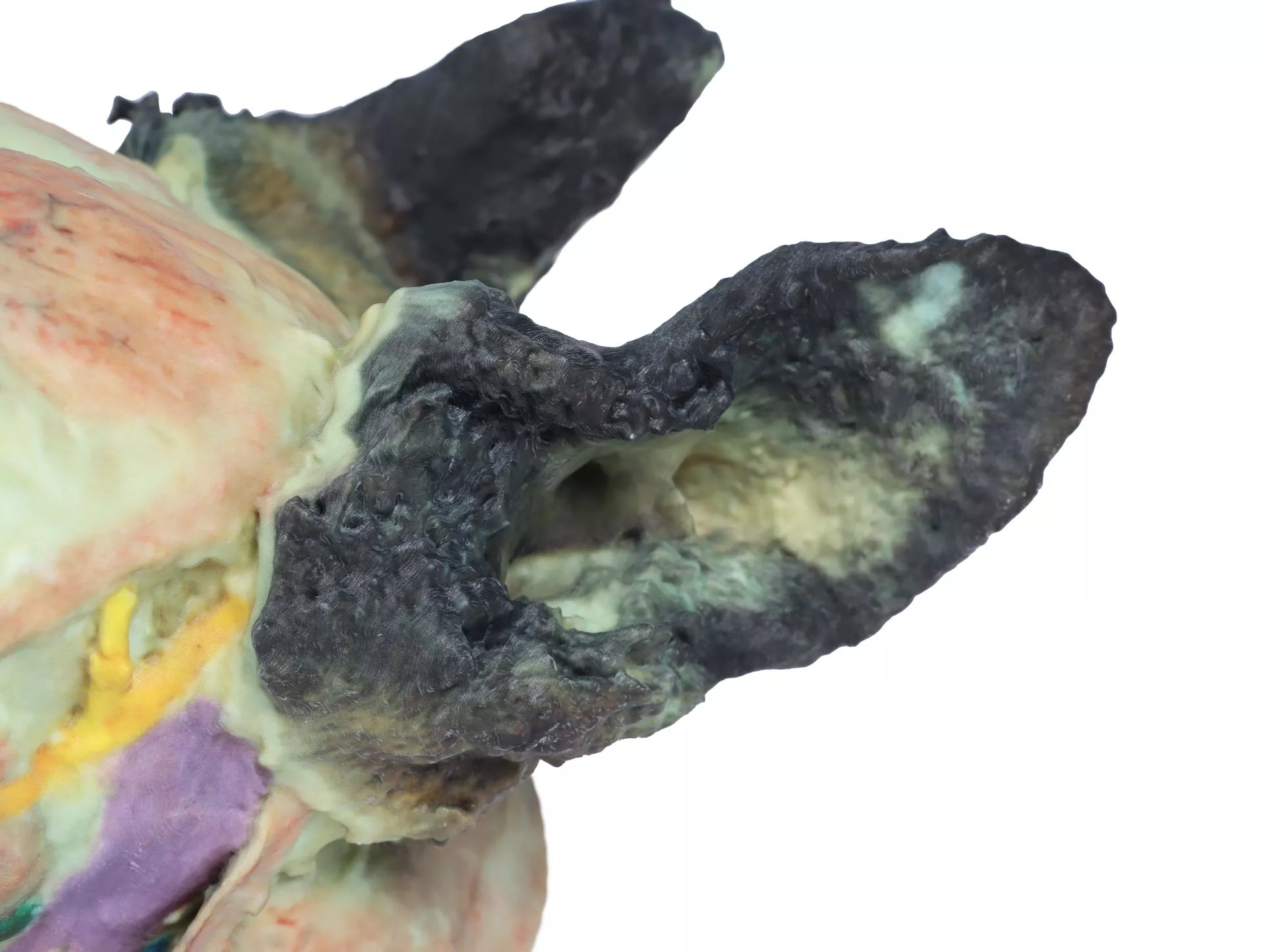
The Dog head – superficial and deep dissections is a clinical-grade anatomical model designed for veterinary educators and medical students. It provides a hands-on reference for studying both surface and internal canine head structures. Choose this advanced model to gain precise insight into neuromuscular, vascular, glandular, and lymphatic anatomy for veterinary teaching or research.
Unlock Comprehensive Canine Anatomy with the Dog head – superficial and deep dissections
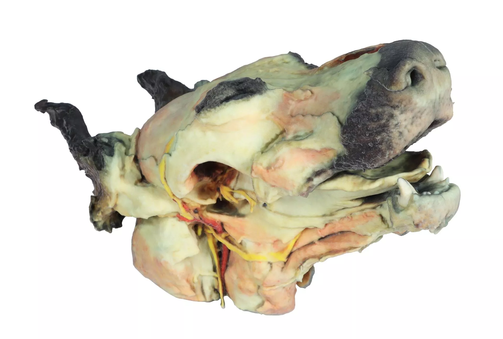
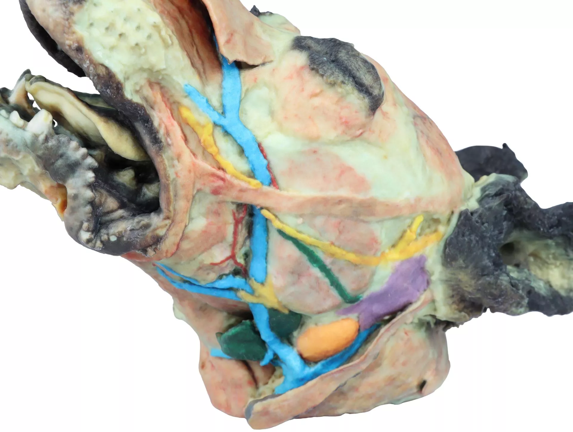
This premium anatomical model features meticulous dissections of the dog head to reveal superficial and deep structures. On the left side, skin removal exposes key facial muscles, nerves, and blood vessels ideal for teaching the fundamentals of neuromuscular and vascular anatomy. The right side offers a deeper dissection with mandible removal, displaying internal muscular structures, major nerves, arteries, and glands. Veterinarians, lecturers, and students benefit from accurate anatomical mapping for improved clinical understanding and teaching effectiveness. With this model, you experience unrivaled visualisation of the temporomandibular joint, oral cavity, and pharyngeal regions.
Features and Benefits
- Shows tip of nose, right and left wings, and non-glandular skin
- Left side dissection highlights major facial and masticatory muscles
- Displays facial, buccal, auriculopalpebral, and mandibular nerves
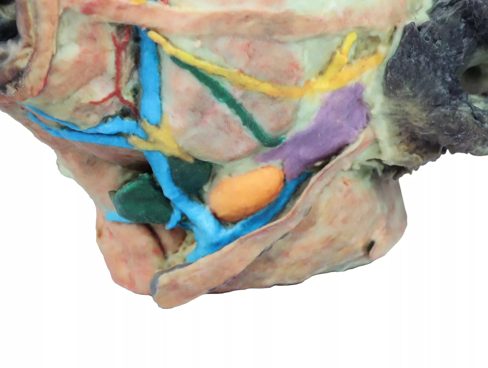
- Exposes facial artery, maxillary artery, external jugular, maxillary, and linguofacial veins
- Identifies parotid and mandibular glands with associated ducts
- Reveals mandibular lymph nodes
- Right side dissection removes mandible for deep structural exposure
- Highlights temporal bone & temporomandibular joint
- Exposes medial pterygoid, styloglossus, genioglossus, geniohyoid muscles
- Identifies hypoglossal and vagosympathetic nerves
- Traces the path of the common carotid artery
- Clinical-grade quality for accuracy and durability
- Perfect for veterinary education and advanced study
Indications for Use
- Veterinary anatomy teaching in academic settings
- Medical student and clinician practical dissection learning
- Demonstrations for neural, vascular, glandular, and muscular canine head structures
- Surgical planning and anatomical reference
Size Guide
- Model represents true-to-size adult dog head anatomy
- Dimensions and precise scale match veterinary teaching standards