Features
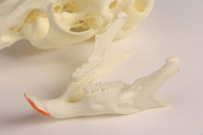
The Chinchilla Skull with pathology, 2-times life size is a meticulously detailed 3D printed model for veterinary professionals, educators, and students. It offers double the real size for enhanced visualization and hands-on learning, clearly highlighting complex dental and jaw pathologies. Choose this advanced anatomical replica to study rare and intricate conditions in chinchilla cranial anatomy with unrivaled accuracy.
Examine Pathological Anatomy with Unmatched Precision and Scale
This model magnifies the chinchilla skull to twice its natural size, making every detail unmistakable for your study or demonstration. Scanned with high-resolution Micro-CT to capture both superficial and internal structures, this teaching tool includes a functional jaw joint and lifelike occlusion to illustrate natural and pathological movement. The Chinchilla Skull with pathology, 2-times life size reveals elongated molars, peak formations, root displacements, periodontal abscesses, and jaw retroposition, allowing for comprehensive clinical education and veterinary diagnosis training. Crafted for reliability and clarity, this model stands out as a cutting-edge educational resource.
Features and Benefits
- 3D printed from high-resolution Micro-CT scan data
- Twice the actual skull size enhances visibility for teaching
- Realistic anatomical details with all superficial structures
- Physiological occlusion for natural bite simulation
- Working jaw joint enables demonstration of movement
- Displays elongated molars and peak formation
- Shows retrograde root displacement and jaw retroposition
- Illustrates periodontal abscess pathology
Indications for Use
- Veterinary education and teaching demonstrations
- Clinical reference for chinchilla cranial and dental pathologies
- Hands-on training for dental and maxillofacial procedures
- Reference model for anatomical research and study
Size Guide
- Scaled at 2-times the life size of a real chinchilla skull
- Exact anatomical proportions preserved at enlarged dimensions